More on birefringence and joining the dots with blood anomalies.
Reference to birefringent technologies that show the same light dispersion details and arrangement using polarized light microscopy.
Above: Cellulose polymerized Graphene nanofibril from a study called ‘Aligning cellulose nanofibril dispersions for tougher fibers’.
Above: sample taken from blood work on an unvaccinated individual, the colours can be reversed by simply adjusting the orientation of one of the polarized filters. These colours can be shown exactly as in the paper reference images linked above, in every detail. Fibers we see vary greatly of course. Many different kinds of nano fiber technology may have been implemented here to support a whole range of biotechnological possibilities.
Regarding only the fibers for now and their details here is the work been done finding possible matches for other criteria rather than using visual techniques such as plain dark field and bright field microscopy. We already know from various sources that analysis has suggested Graphene, Polymer, and other exotic materials to be present. Many images have been taken using a special DIC polarized light microscope during this form observation venture.
These images have highlighted details within fiber, gel like structures, crystals, and more. There are several possible theories on some of these structures, most involving liquid crystals, polymers, and materials not of nature such as part Graphene compositions. It does seem like an interesting variety of artifacts are present and that most of these suspicious structures in question are indeed exhibiting birefringence. But not all. There is still a heavy interest in the organism side of this complicated relationship within the suspected synthetic biology and with other phenomena seen occurring on the samples slides. Most of these visual exhibitions are starting to be realized as a common visual occurrence in the detail of most blood slides and work seen from other sources. Particularly in fact the greenish lipids which attract such biological type attention when using time lapse video analysis. These lipds may be part organism based as discussed before. Maybe there is a connection here to what Clifford Carnicom is explaining to us. These rather different representations observed using completely different analysis techniques need to be aligned, or shared with detailed explanation. The correlations must be made in relation to anything that might match these points in scientific literature. Much literature is already appearing that gives light to the direction we likely should be heading in.
Either way, the implications of any or all papers read so far are that something extremely complex is occurring and likely in tandem with other separate technological advances in order for these structures to be of any practical use.
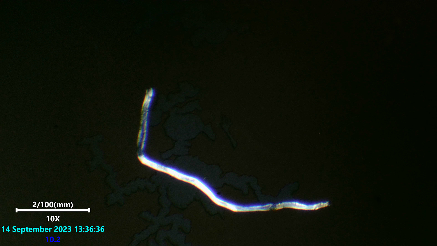
There are the occasional structures which exhibit much less interesting colour waves. These are possibly intended for different purpose, or have not been in proximity to necessary materials in order to form as intended.
Above: a polyester fiber exhibiting less detail or colour wave complexity than seen in blood grown or environmental fibers.
No fibers matching the same light scattering patterns or colour wave details has been seen to reference in literature yet. A closer visual match is has been made to nano technology related paper image references. The fact these are almost 100% of part polymer nature is evident from all literature and image references alone so far. I am inclined to trust some of the analysis done by others regarding these materials and find this form of polarized light microscopy technique to only enforce this likelihood.
The Question is now……..What else is embedded within these fibers and structures that make there light wave patterns and visual formations vary so much from simpler and less nefarious technology forms ?
More on this to come.
A thank you again to those helping with small contributions or equipment.


