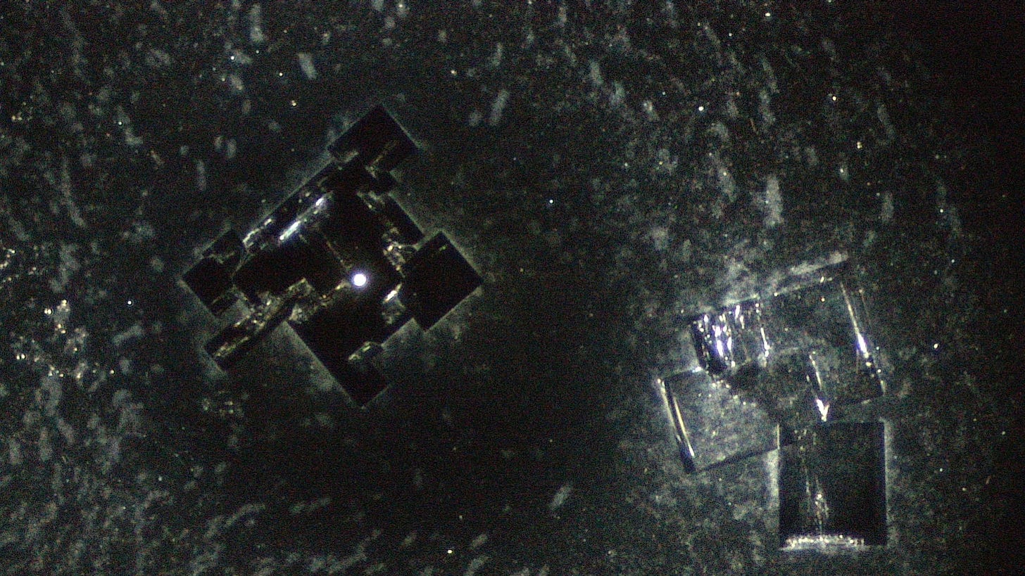
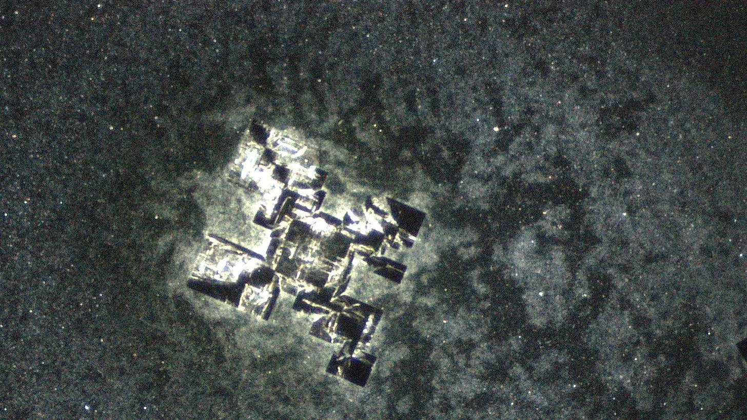
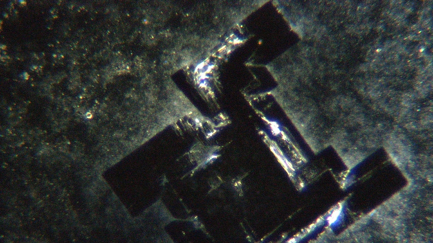
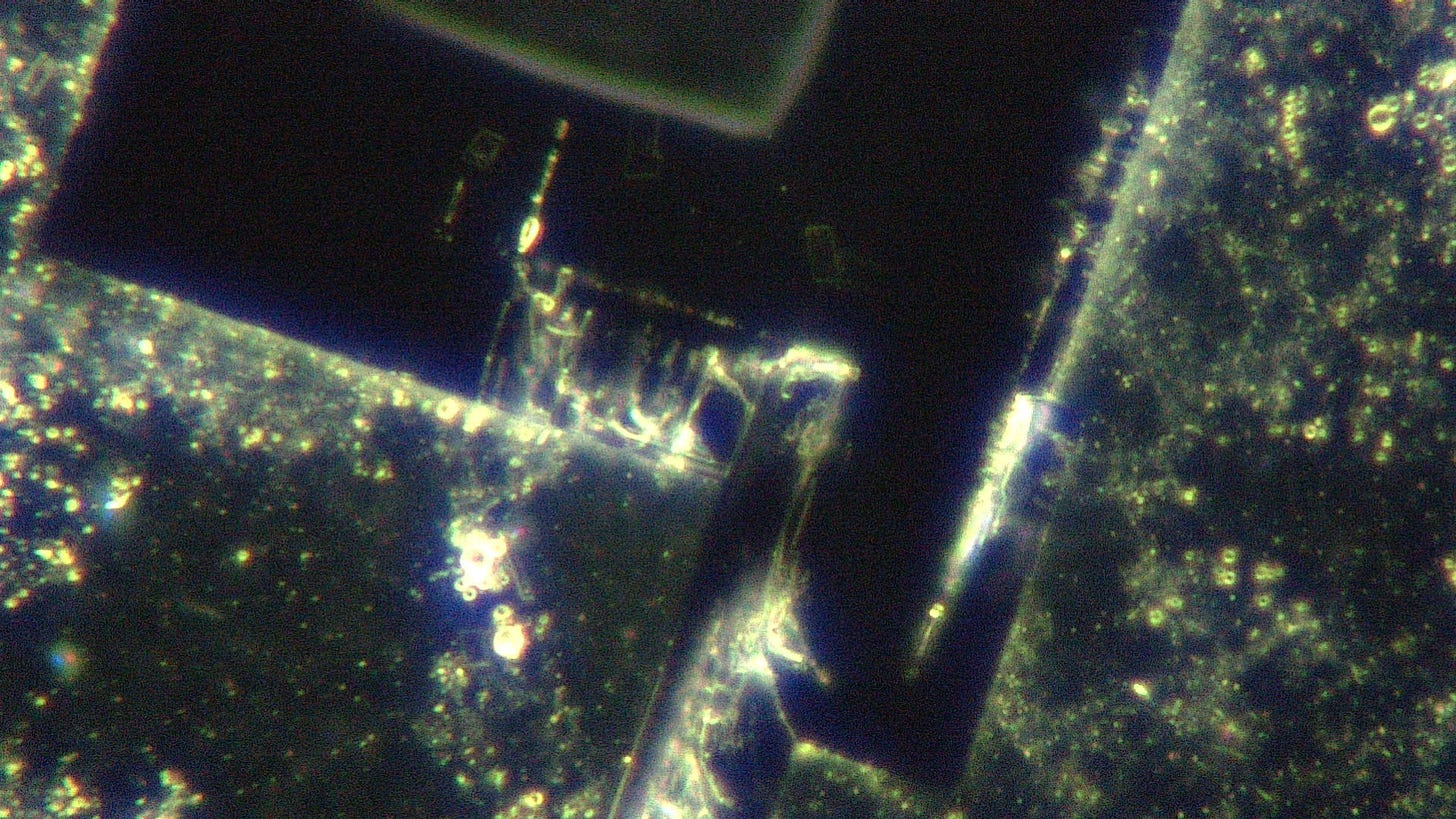
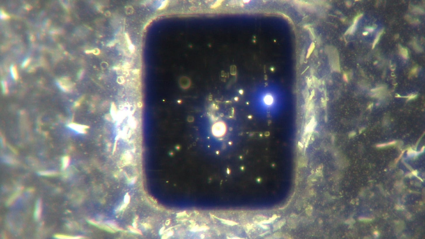
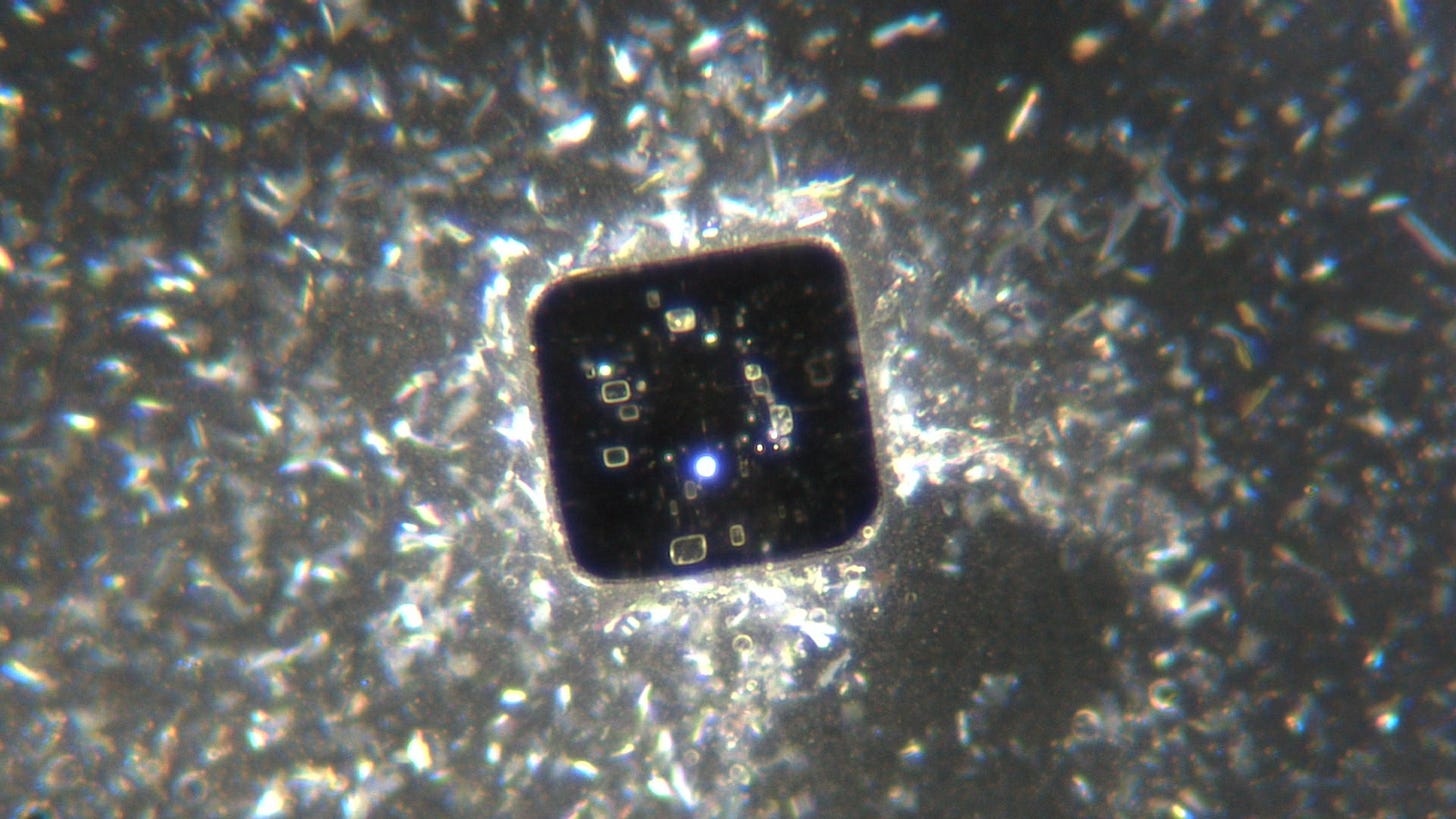
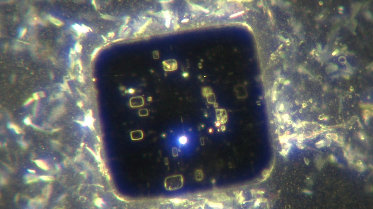
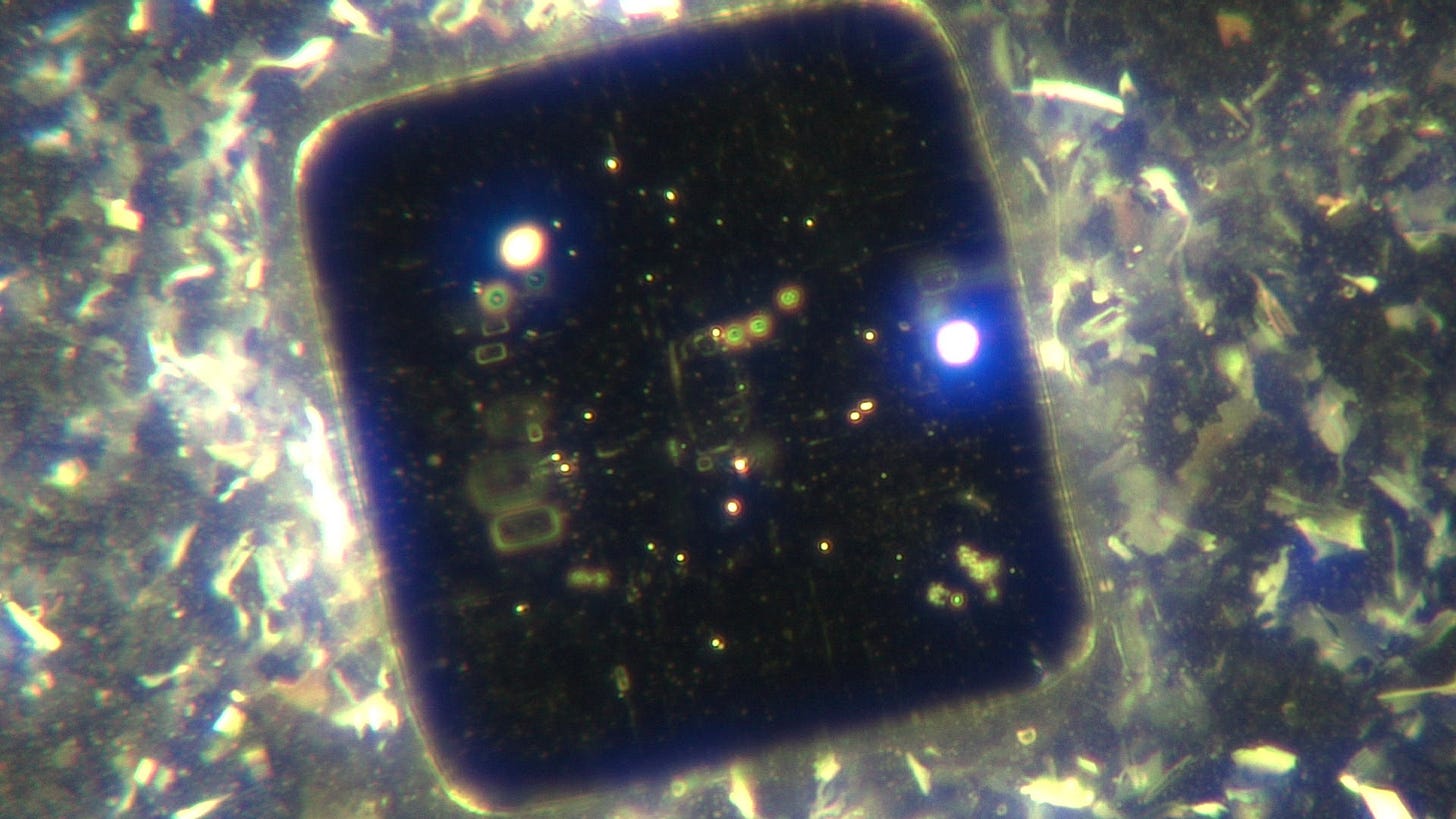
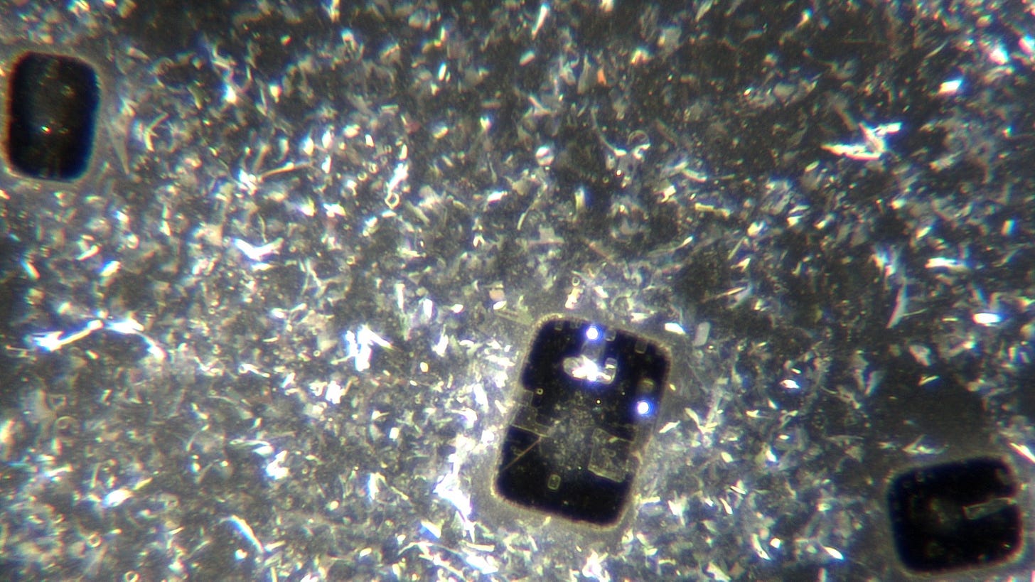
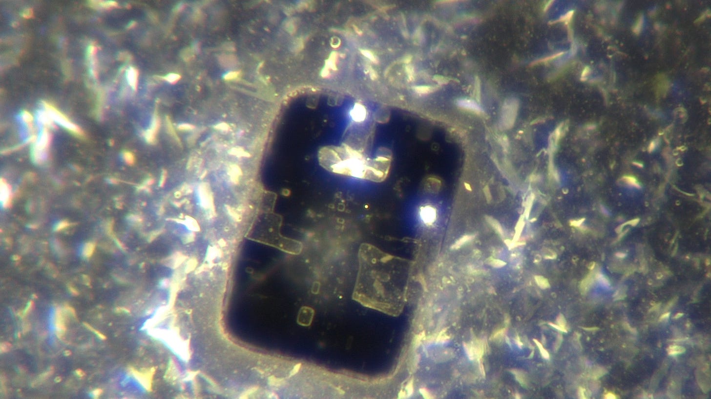
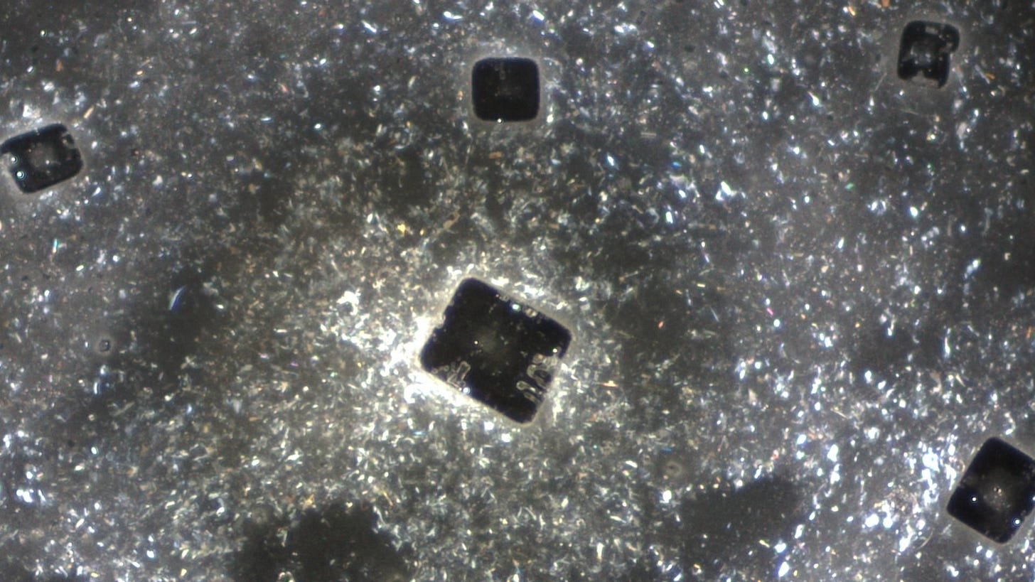
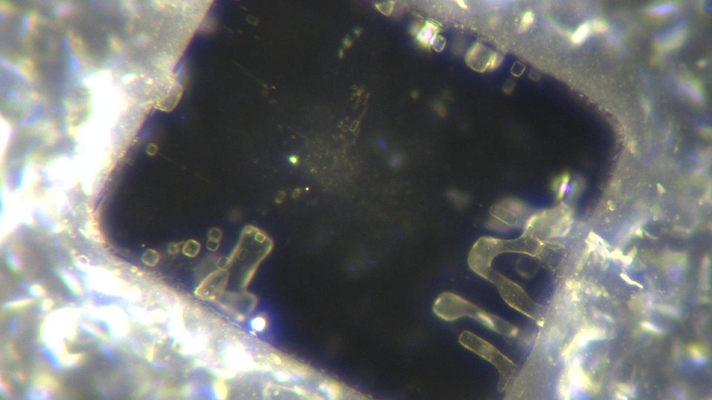
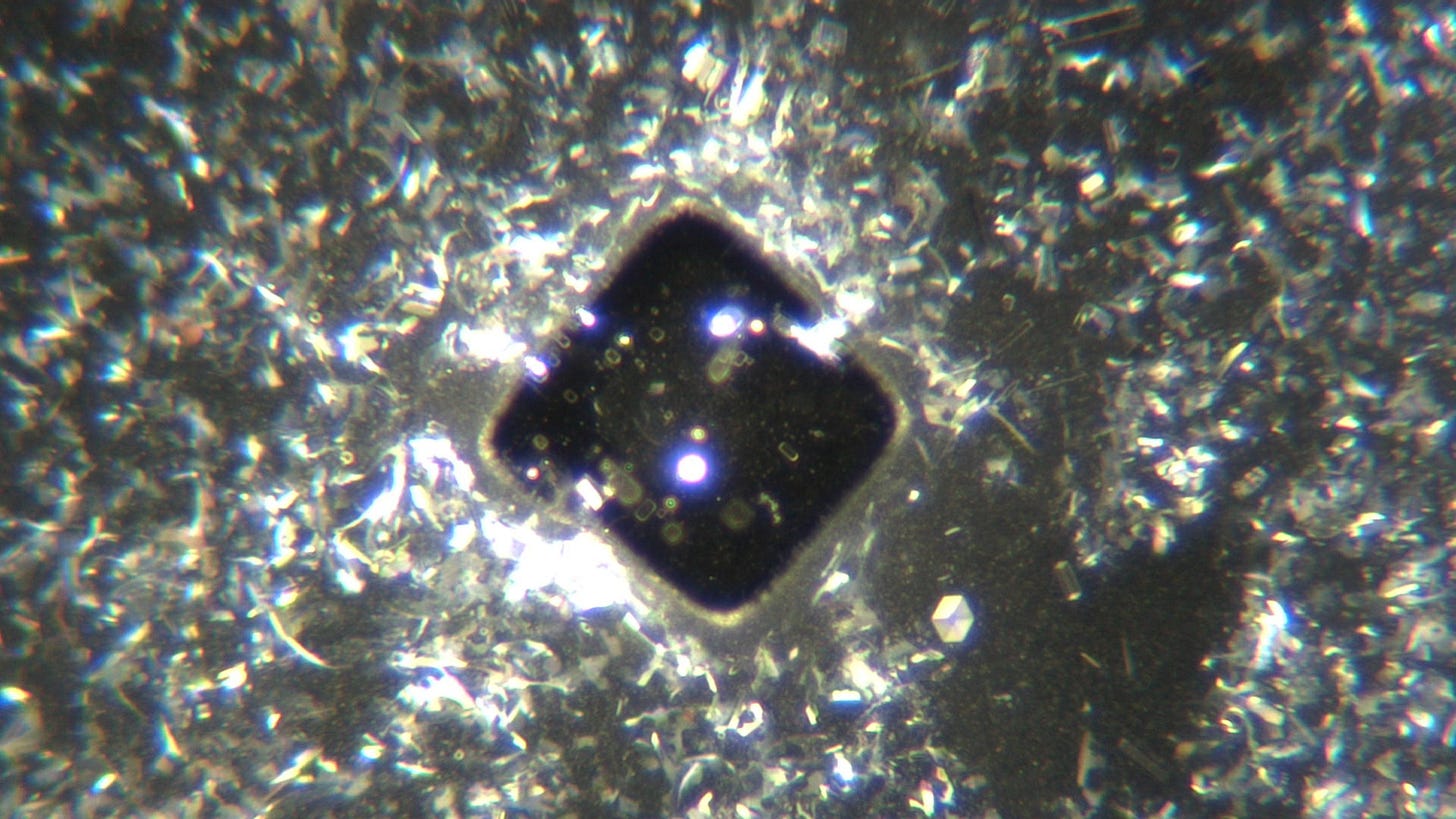
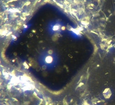
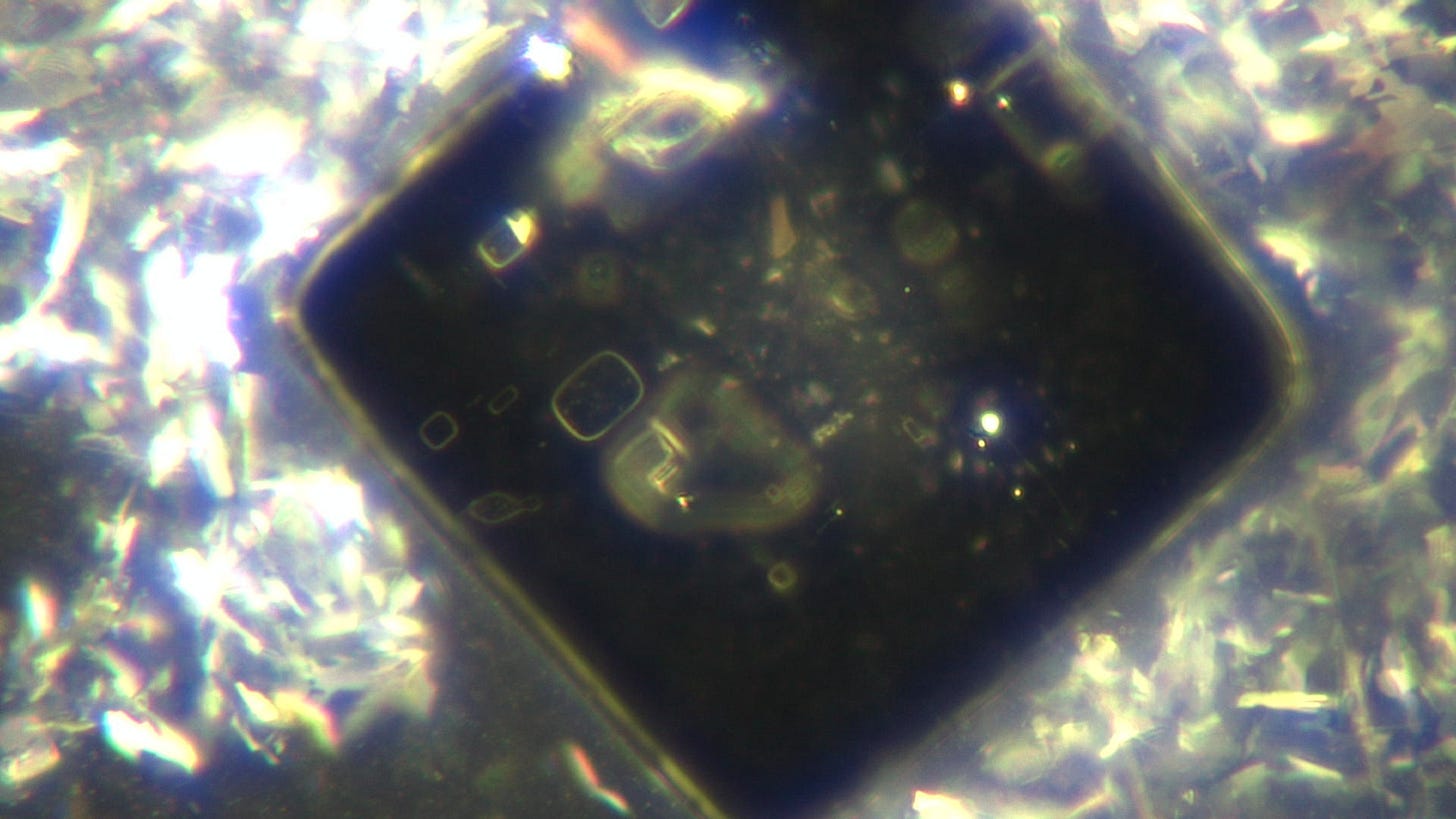
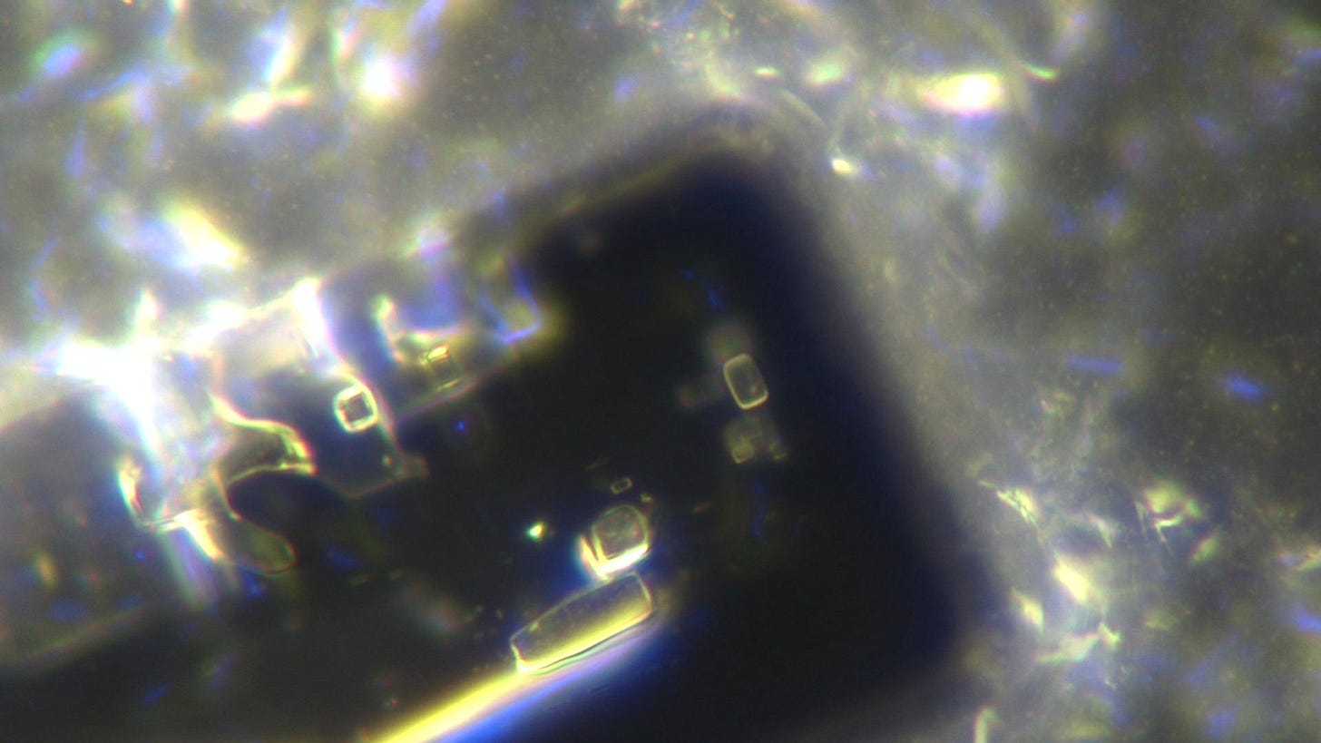
Comirnaty sample from August 5th 2022
(from a more concentrated sample - similarities and differences)
On the 5th August I prepared two slides in a similar fashion to that described in my last couple of posts with one potentially important difference. I concentrated the sample by means of a centrifuge - from memory at about 2500 rpm for 15 mins. I then took my samples from the bottom of this test tube.
I placed a few drops of this concentrated Comirnaty on a slide and allowed the sample to partially dry before adding further drops. I am fairly sure I prepared a second slide after I placed a coverslip on the first sample in order to get a closer look at a large structure (see below). The time stamps of the images suggest this too.
All these videos are in real time, dark field and either at 40x magnification, or at 100x or 200x magnification.
In this first video one can see the ‘usual’ sparkly material that has no place in a pharmaceutical product, as well as larger metallic looking objects that float past. The drying pattern which starts in the top right corner includes some particularly straight patterns - just an observation eh…
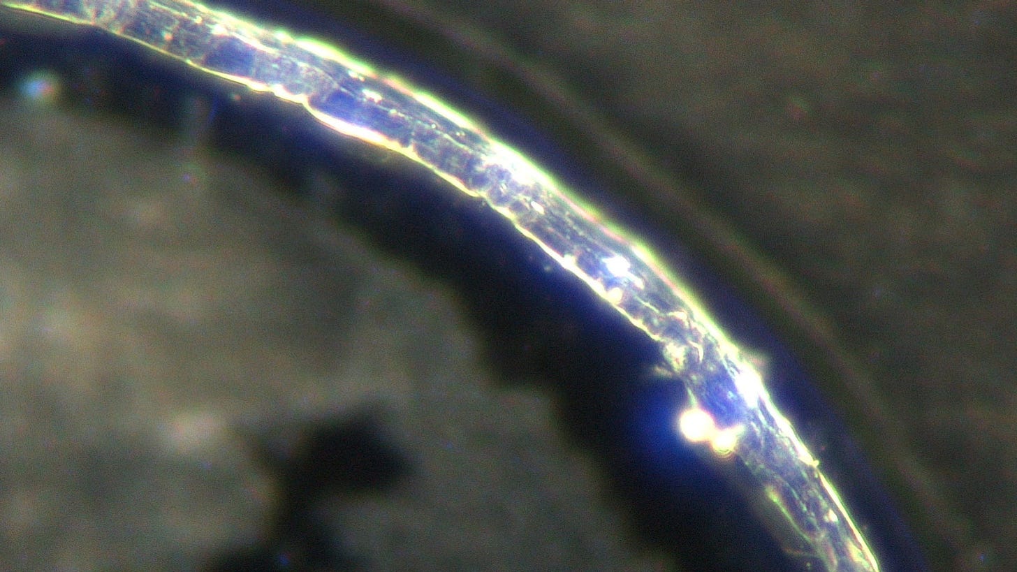
In this video check out the appearance of a typical ribbon structure at the 5min mark:
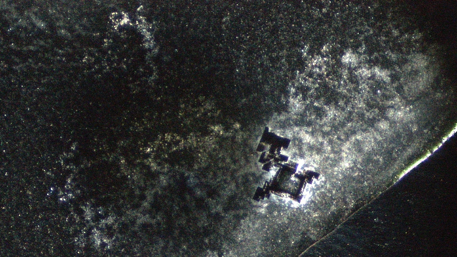
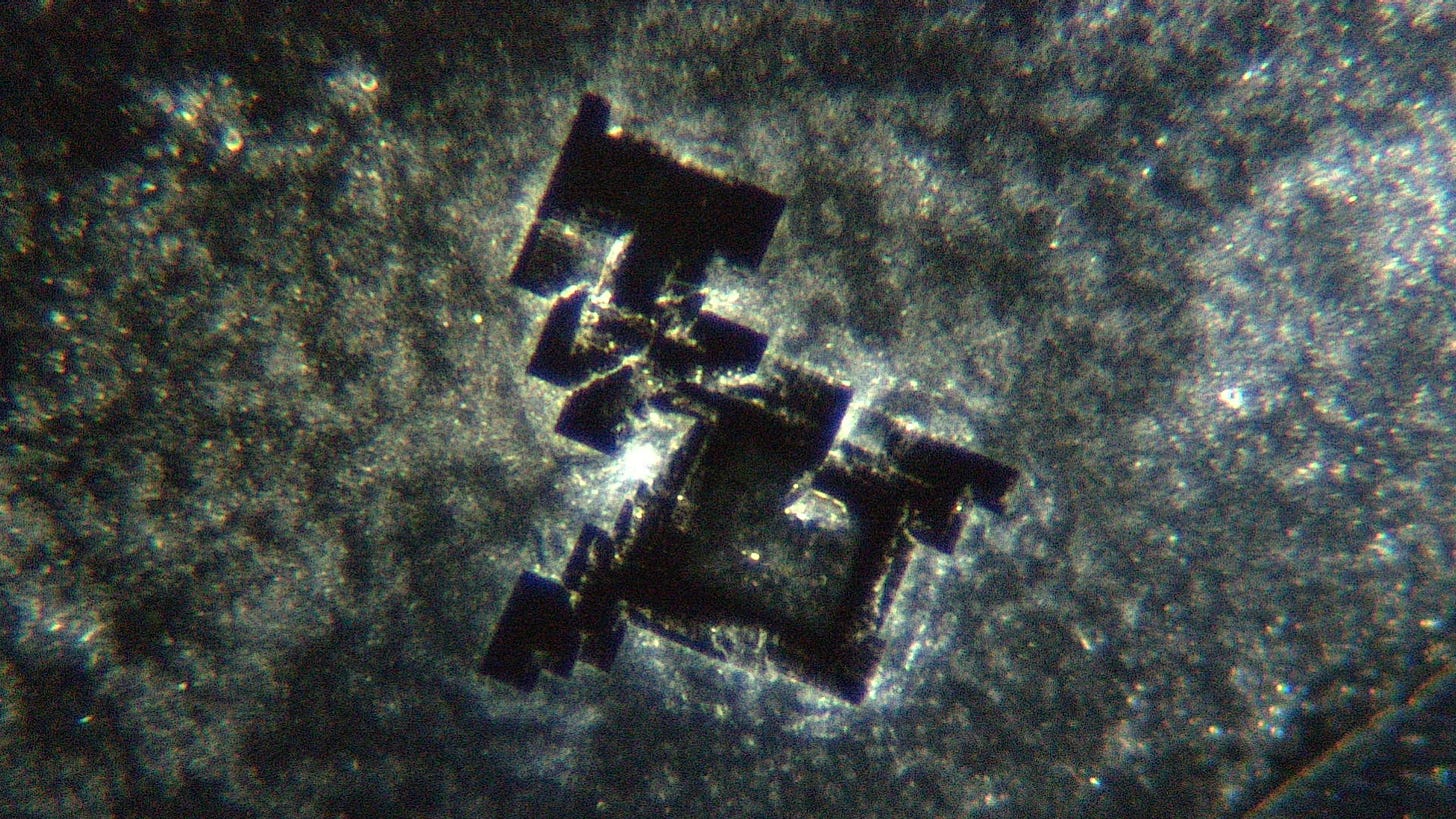
This very bright structure was most dramatic
40x
200x
1000x and under a coverslip, using oil immersion in the hope that I could get a closer look, which I did but rendered the slide unable to be of further use. Further images are from the second slide. I think it was worth it though WTF is this?
There were also a few other ribbons that appeared as the sample dried. This been one of them:
200x:
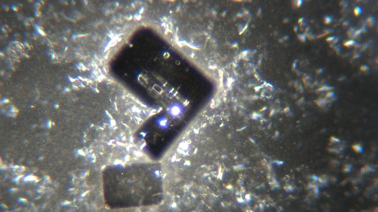
and a few ribbons visible after the sample dried. This one at 40x:
200x:
40x
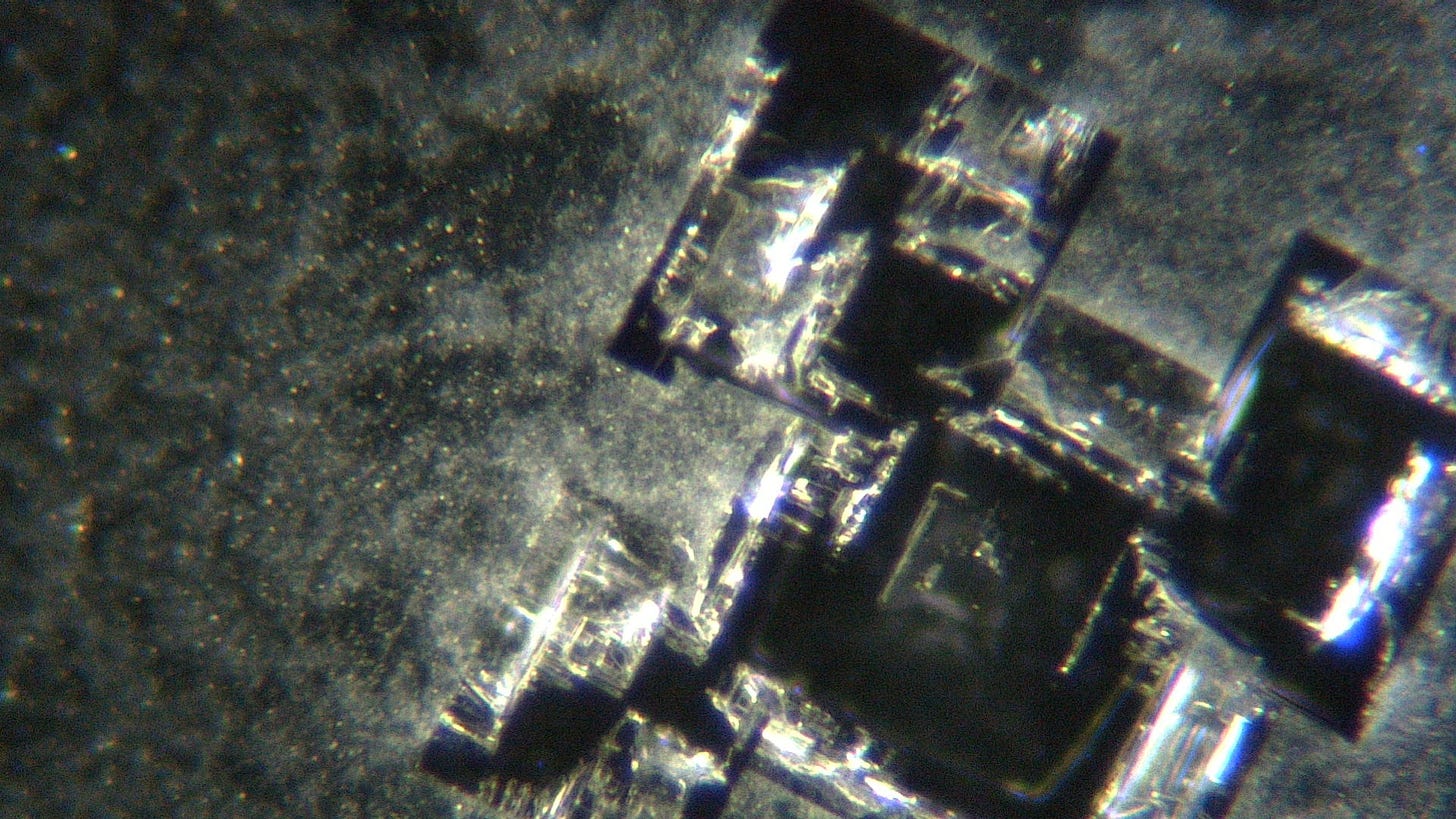
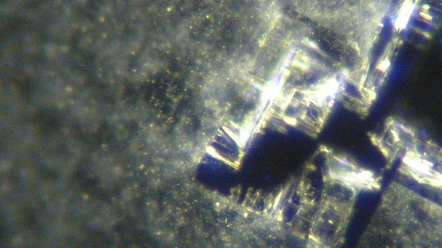
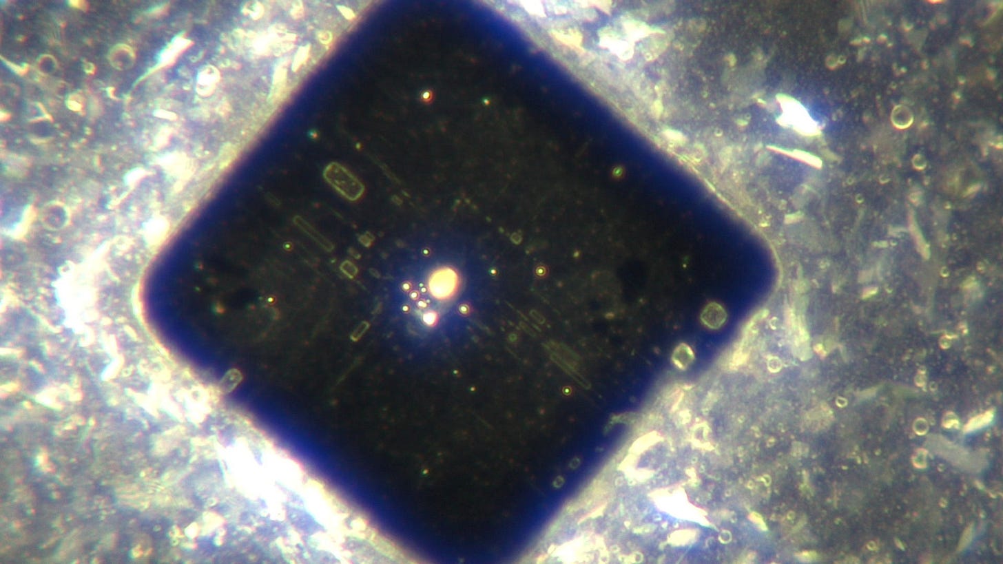
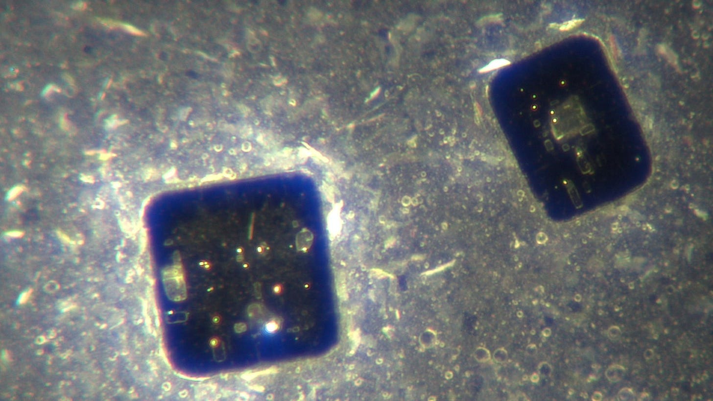
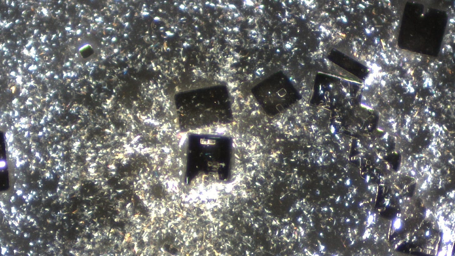
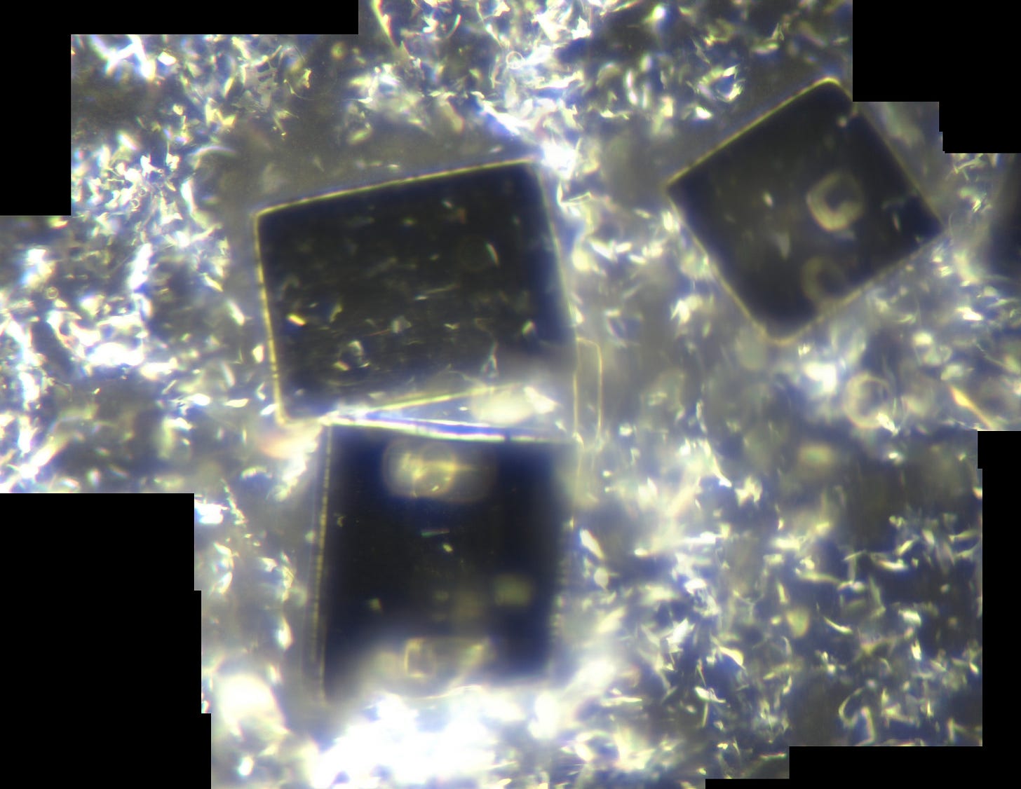
The initial crystals that developed were generally more complex in structure and were photographed the following day i.e. the 6th August 2022:
closer view at 200x magnification of bottom left corner:
another example at 40x magnification:
200x
40x
100x
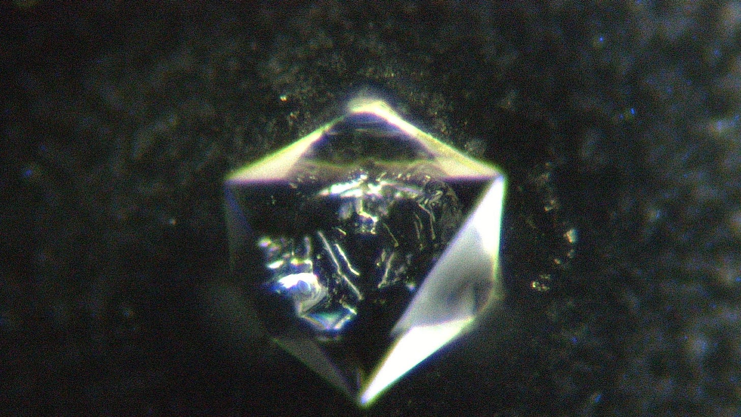
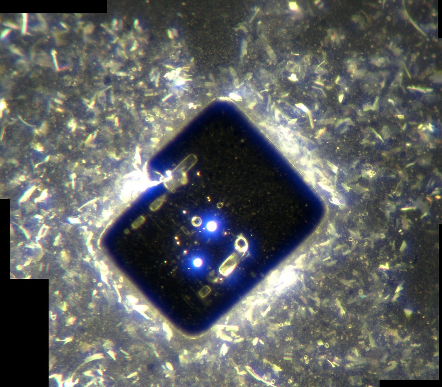
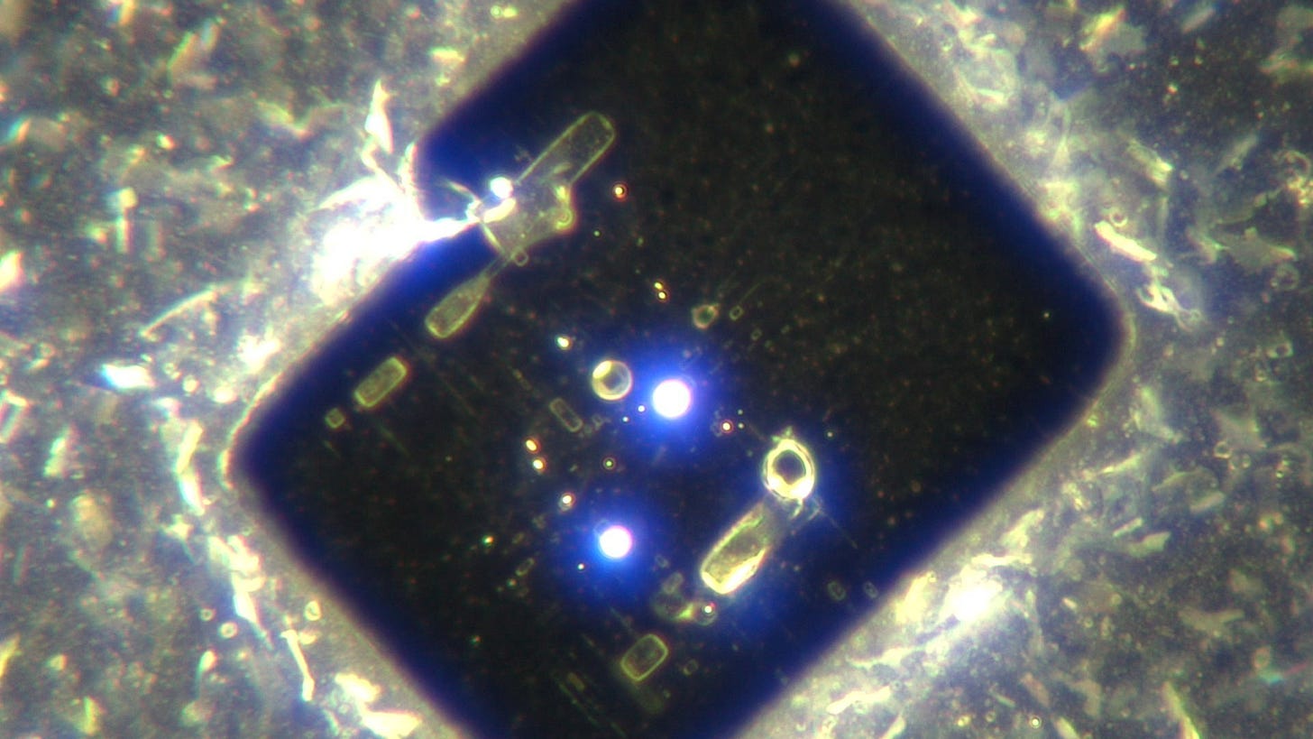
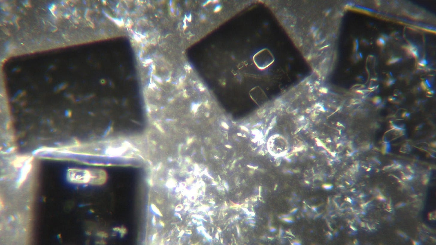
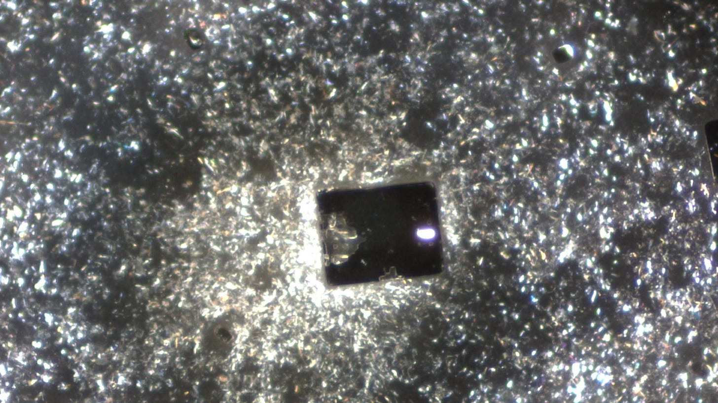
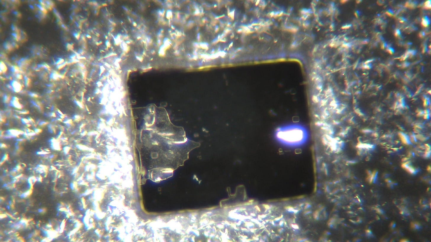
There were also translucent crystals. This one is a beauty:
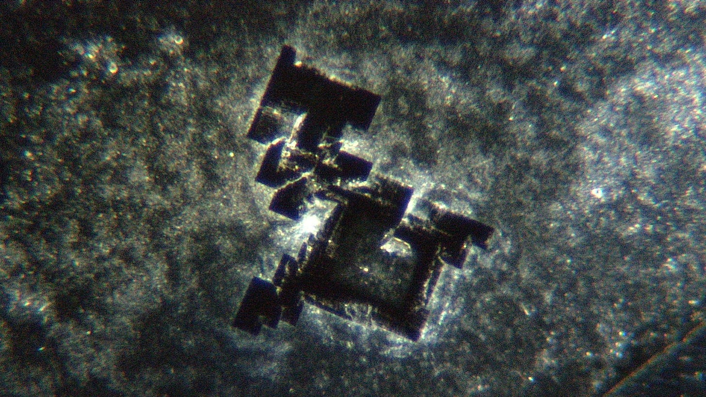
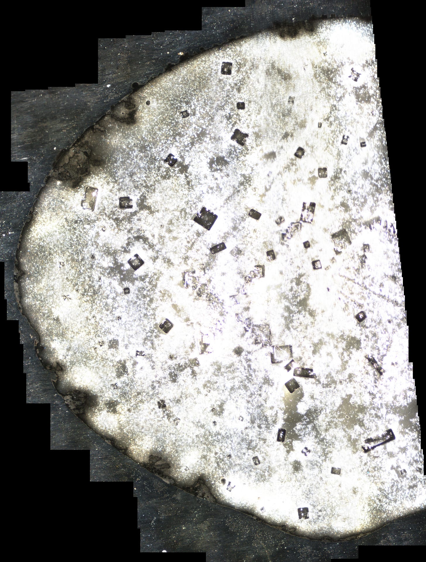
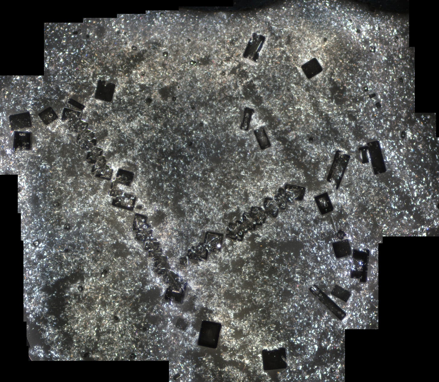
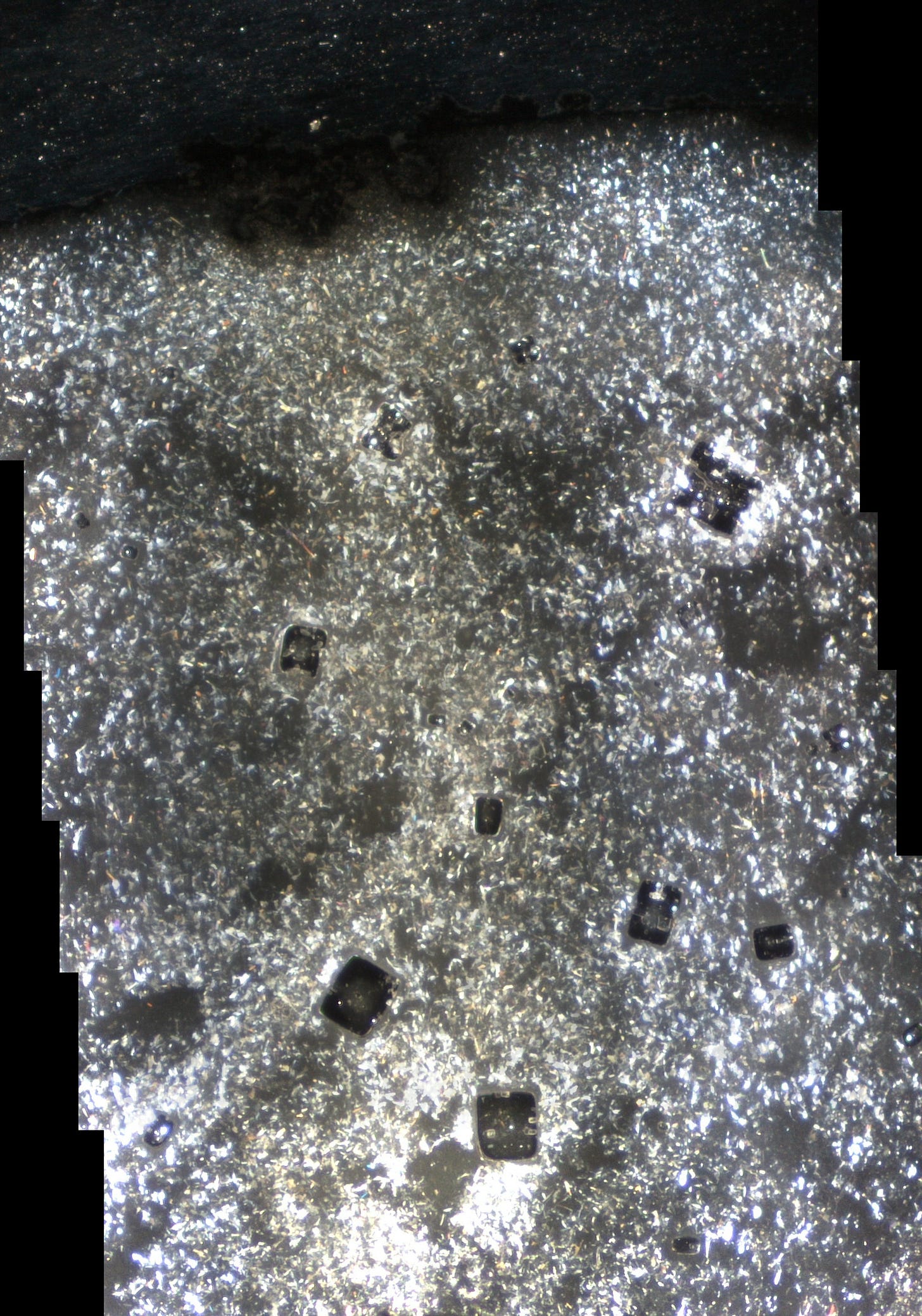
Like the other two slides prepared in early August this slide sat in a drawer for 2 months. These photos are from 20th October 2022. Here is an over exposed photo-stitch of the left hand end of the sample. It does illustrate a few things however. The initial structures are predominantly peripheral whilst the majority of these structures are away from the edges. There structures appear appear at first glance to be positioned haphazardly but having looked at these for a while they are (mostly) in lines and many are associated with the ‘X’ structure centrally. There are also ‘ghost’ squares where experience will tell me the structures used to be present.
fantastic cluster:
closer:
Phew that’s a few eh… The lack of ribbon structures seen in the liquid suggest these structures are from a second sample. But maybe there is another explanation. The background ‘gel’ is also brighter, and as Karl C has pointed out the structures have rounder edges too. Maybe the ribbons don’t form because the conditions are different. Ron Norris has commented recently that the gel can change state from a gel to a fibre depending on pH. Perhaps the concentrated sample varies enough in some respect that the fibres don’t form?? And that condition also changes the form of the structure? Some of the surface details of the structures are different too. Are they less well formed or better formed?
I look forward to further discussions.
Hope Y’all have had a great week!
David


