Masterpiece of Deception- A Trilogy
Part 1: Laboratory and Microscopy Investigation into a Zeolite Product
The internet and social media are going crazy about a new product on the market that contains -according to the manufacturers - nanosized zeolite of the type clinoptilolite. It’s being hailed as a panacea for removing the nanotechnology injected under the ruse of Covid vaccines, but we’re also exposed to via chemtrails food and water.
It’s not my intention to destroy products and companies and I have no gain from doing that. However, I’m after the truth and if I see that a product can cause more harm than good, I will speak out about it after having done the proper research.
I first heard about this product from a friend abroad. When I first looked into it superficially my thoughts were, well it all sounds good, but where is the evidence? Testimonials are not enough for me to come to a conclusion if a product works as the placebo effect is very powerful. For example, a study on the effectiveness of SSRI antidepressant medication found that they only work in 20% of patients as 80% react just as good when on a placebo. Then, another friend abroad told me about a discussion he had with them and a study the company put out. That’s when I began looking more closely at this product. I will dissect the study in part three of this trilogy.
What is Zeolite?
There exists naturally derived Zeolite and synthetic Zeolite crystals. Zeolites classification can go beyond the origin though and be based on their adapted structures of which up to now 200 types of synthetic zeolite structures and 10 structure types of natural zeolite have been discovered.
Chemically Zeolites are crystalline, hydrated aluminosilicates. This means they are made from Aluminum, Silicon and Oxygen. Aluminum activates polymerization (self-assembly). Silicon is used in nanotechnology and computer chips. I mentioned aluminosilicates in my article showing microscopy of various liquid medicines and supplements. See here:
https://anitabaxasmd.substack.com/p/microscopy-of-edta-glutathione-ivermectin?r=1sq8ta
Specifically, glass vials made by Corning used for injectables are made from aluminosilicates. Aluminosilicates are defined as minerals used in drug delivery systems due to their porous structure, biocompatibility, and ability to entrap and release drug molecules efficiently. This means drugs can be inserted into them to be delivered to different parts of the body.
Areas of research into this product
Since the manufacturers state that it’s made from marine plasma, I suspected it may contain some toxic metals. I sent it to a lab I have worked with for many decades to analyze it. Next, I did bright- and dark field microscopy on it. I exposed samples to constant EMF by placing them on a wireless phone charger for up to 12 days and re-examined them to see if something changed or rather if something grew.
I added it to the blood of a severely ill jabbed person to see if it made any difference. Besides this practical testing, I researched articles and studies showing that Zeolite is used in combination with polymers and Graphene oxide to enhance the abilities of nano-sized bio sensors. I also looked at the study on the website of the manufacturer done with three (only 3!) people supposedly showing a reduction of Graphene oxide and Aluminum. I am presenting the findings of my research in three parts over the next few weeks. So, let’s begin with what I found in the lab results and with microscopy.
Toxic Metal Analysis
Since the lab doesn’t analyze products, but only hair, urine, stool and blood I had to use a bit of subterfuge to get the analysis done. I took a urine sample and filled one sample container of 40 ml with only urine and the second one I filled half with the urine of the same sample and half with that product.
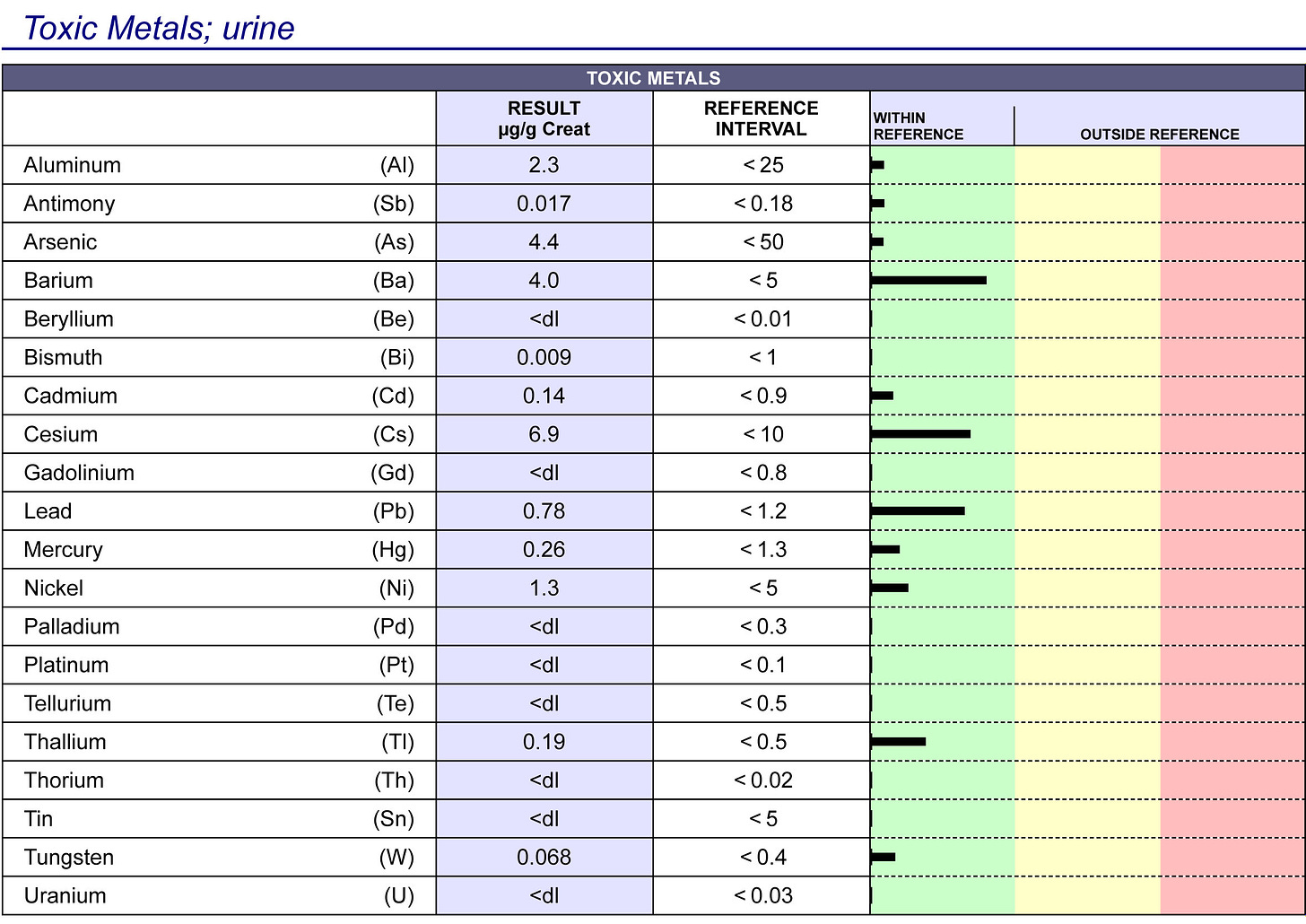
This is the analysis of toxic metals in urine only: It shows low levels of Aluminum, Antimony, Arsenic a bit more Barium, Cesium and Lead and again lower levels of Mercury, Nickel, Thallium and Tungsten all within so-called normal range (green area).
Above the results of the pure urine sample
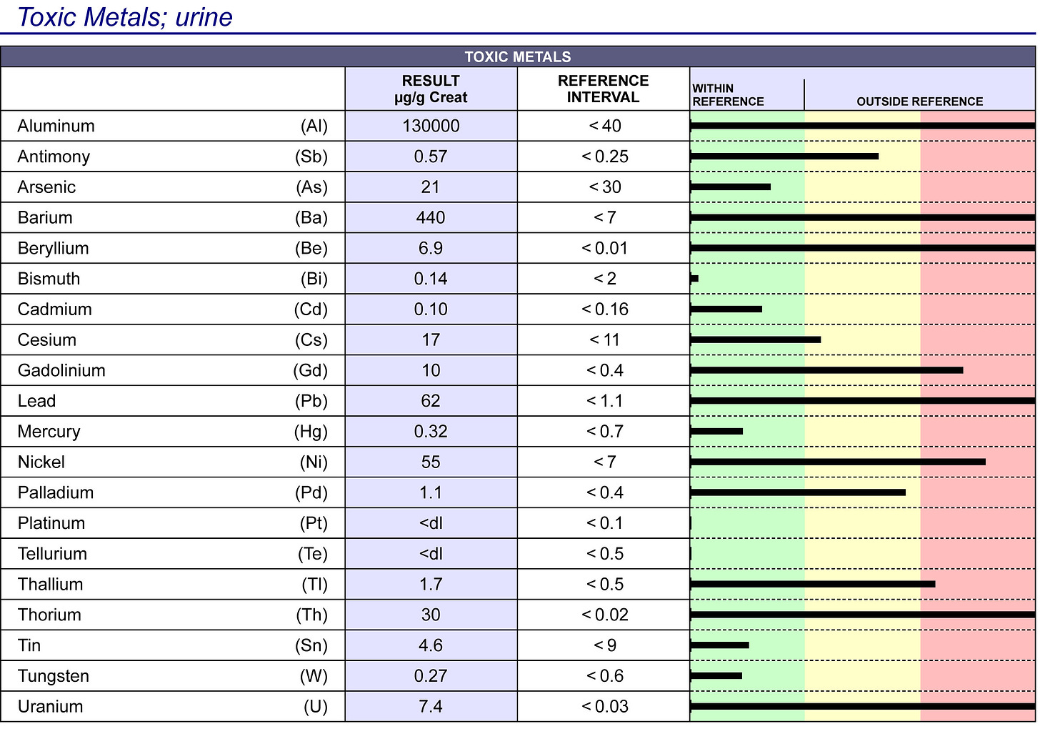
My hunch was right. The product contains many toxic metals.
Following is the analysis of the sample containing half urine of the same urine sample as above and half of the product. The toxic metals are literally off the chart. There are metals that were not present in urine alone such as Beryllium, Gadolinium, Palladium, Thorium and Uranium. Note that the “normal” value or reference interval for the sample below is higher than for the sample above. This makes these results even worse as higher amounts are “allowed as normal. “
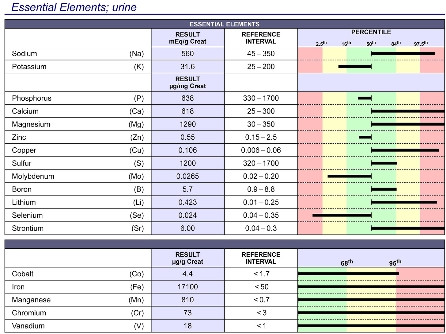
Essential Elements Analysis
The manufacturers state that their product contains many minerals because it’s made from marine plasma.
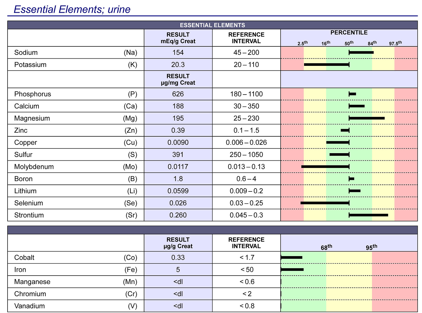
Below are the minerals found in the pure urine sample to compare with what was found in the urine/product mix.
Below are the minerals found in the half and half mix.
The product does contain minerals as advertized as the values are higher than in urine alone for Sodium, Potassium, Phosphorus, Calcium, Magnesium, Copper, Sulfur, Boron, Lithium and Strontium. It contains a tiny bit of Zinc and Molybdenum. It contains a lot of Cobalt, Iron, Manganese, Chromium and Vanadium.
Testing Methods
The toxic metal analysis was done by ICP-MS triple quadrupole (QQQ) in a laboratory that has been doing such testing for decades and is one of the world's leading laboratories. An ICP-MS triple quadrupole (QQQ) testing method is a type of Inductively Coupled Plasma Mass Spectrometry that utilizes three mass analyzers (quadrupoles) in tandem, allowing for enhanced selectivity and sensitivity by isolating specific ions through a collision/reaction cell.
Essential elements testing was done with ISE (Ion-Selective Electrodes ) and spectrophotometry. ISEs are electrochemical sensors that use a specific membrane for each ion to measure electrolyte activity. ISEs are considered the most accurate method for measuring electrolytes. Spectrophotometry is a technique that measures the color produced by radiated light. Spectrophotometers are also known as UV-Vis spectrometers. The output of a spectrophotometer is usually measured in the absorption spectrum of the sample. The results are reported as micrograms/g creatinine. Since the sample with the product was diluted with urine, the creatinine level obviously was lower. The pure urine sample had a creatinine of 51.4 mg/dl and the product/urine mix had a creatinine of 20.7 mg/dl. Therefore you can divide the toxic metal results by 2.48 (51.4 : 20.7) and you still get enormously high toxic metal values. For example, Barium in the mix shows 440 micrograms/g. Divide this by 2.48 and you get 177.4 micrograms/g creatinine. The pure urine sample contained only 4 micrograms/g. The mix contains 44 times more Barium than the pure urine sample. You can calculate this with the other toxic elements you will find the levels of the urine/product mix are extremely high. There are metals in the mix that do not appear at all in the pure urine sample such as Beryllium, Gadolinium, Palladium and others.
Microscopy Findings
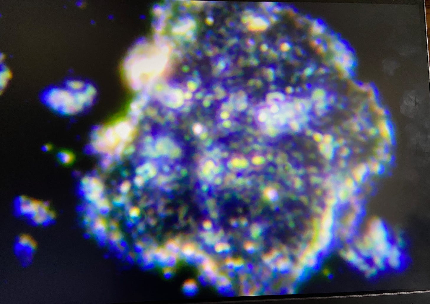
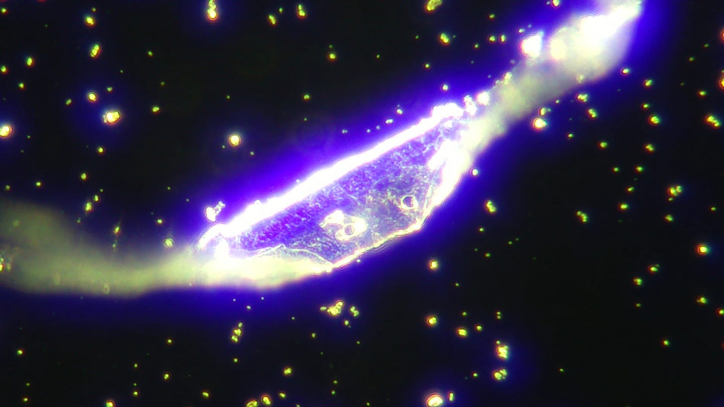
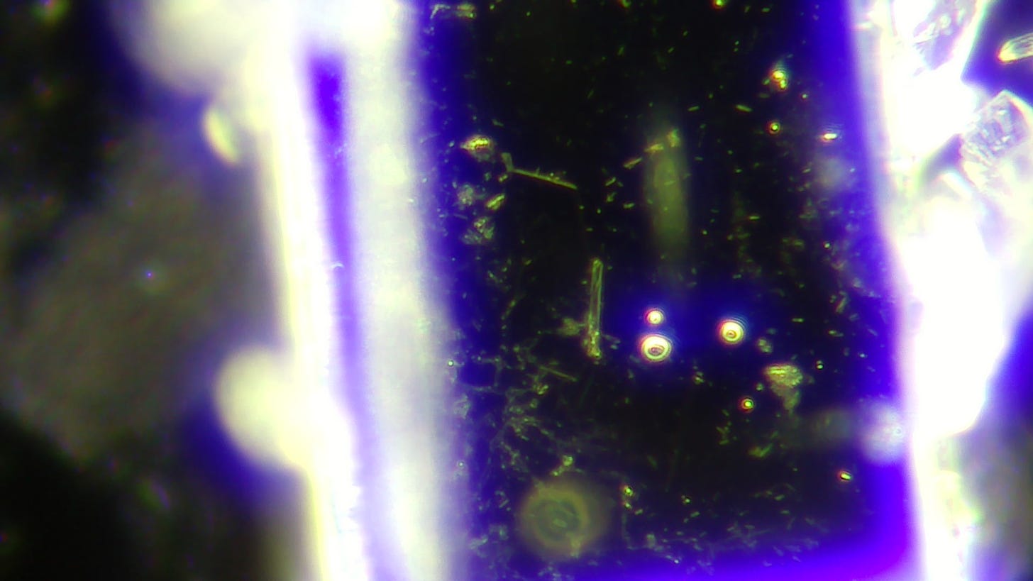
When I took the first look through the microscope in dark field I was taken aback at the blinking and moving things I saw en masse. This video is a good representation of the many times I have seen these moving, blinking things:
It reminded me of the video Dr. Ana-Maria Mihalcea MD, PhD put up showing lots of moving particles in the Pfizer BioNTech vials here:
https://substack.com/home/post/p-150734338?source=queue&autoPlay=false
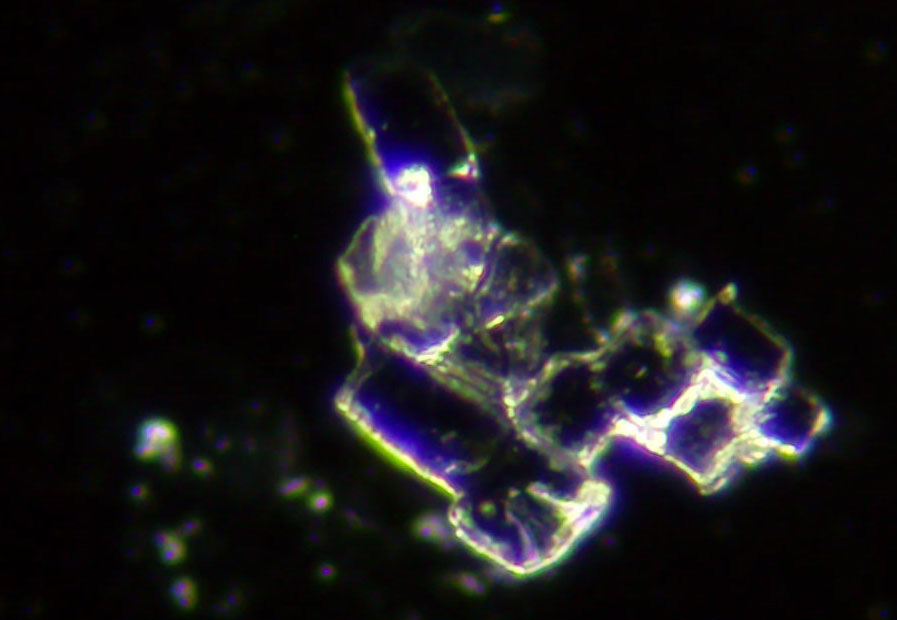
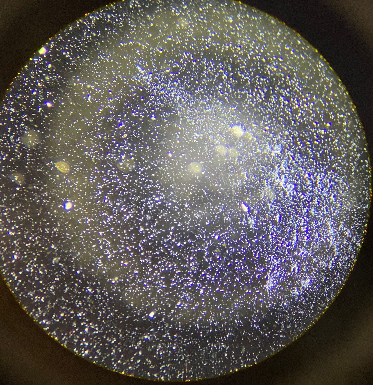
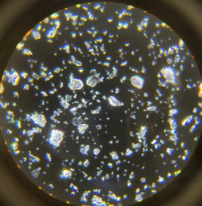
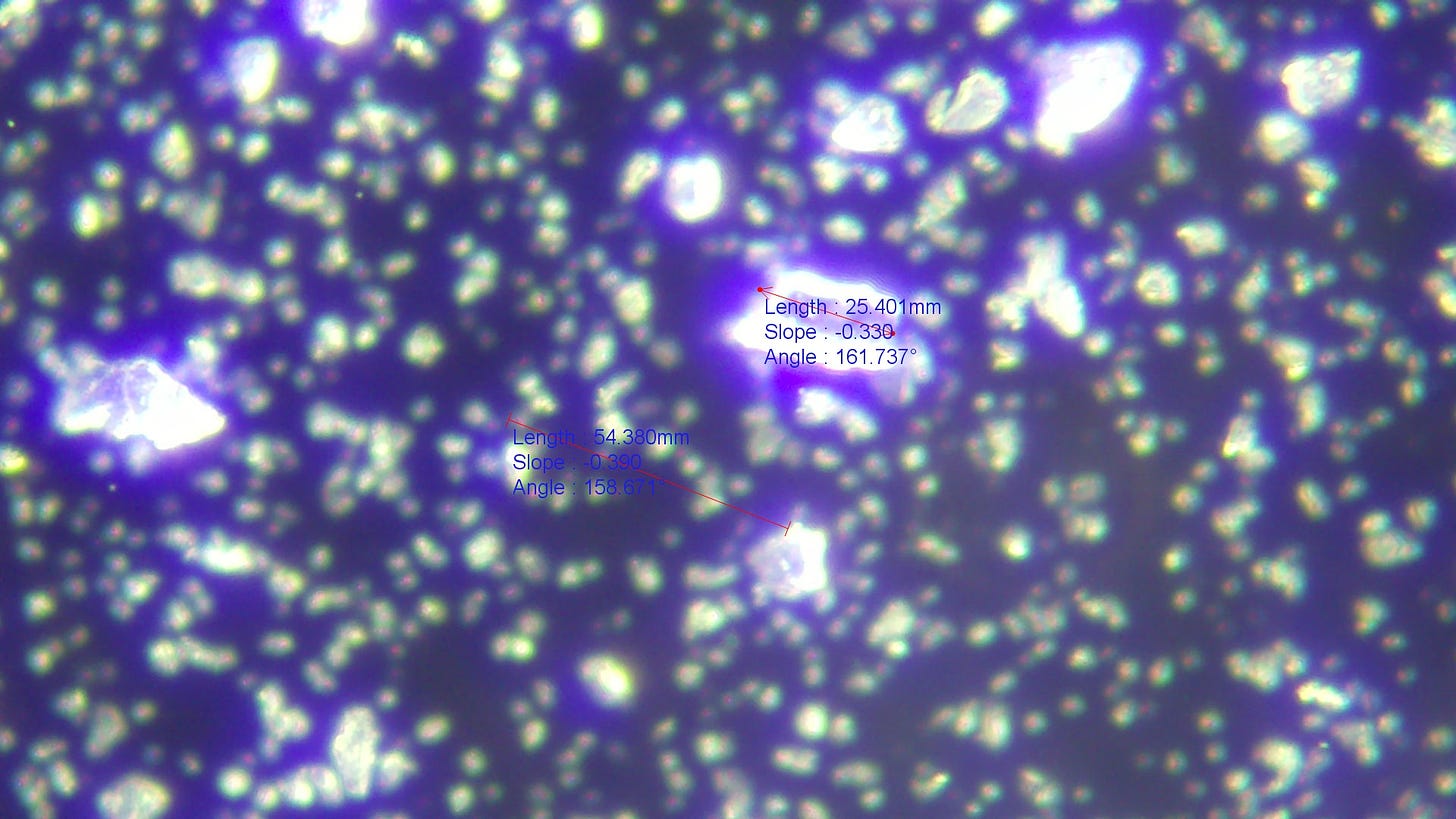
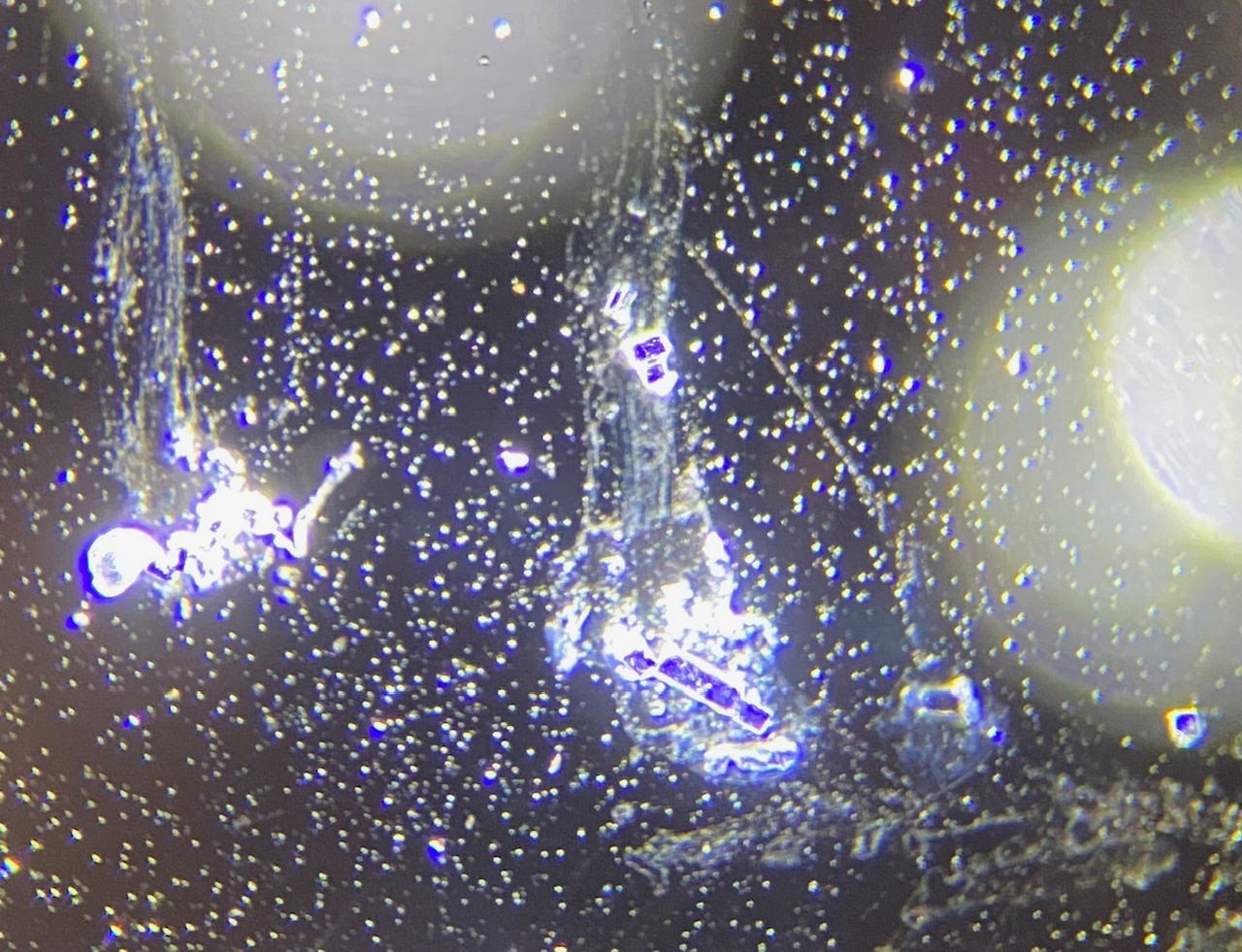
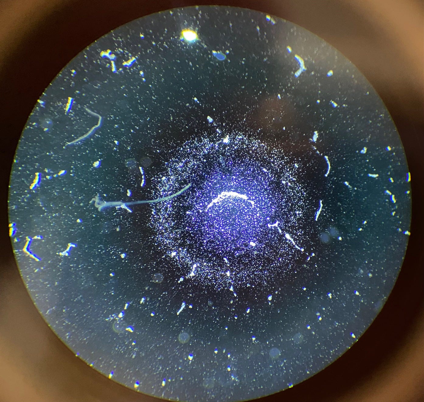
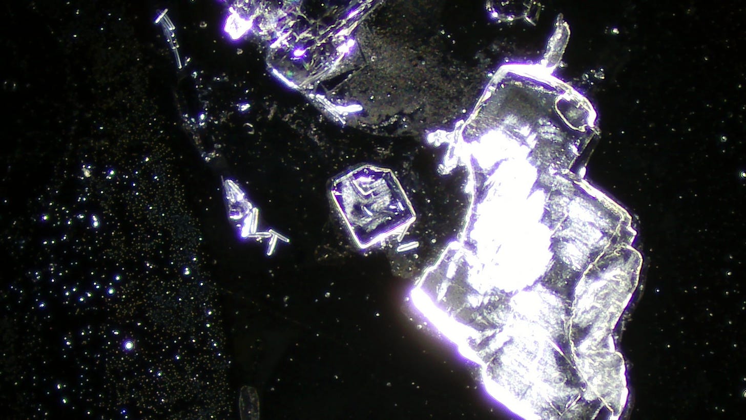
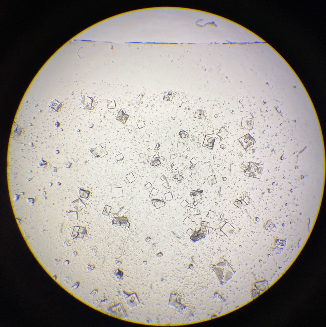
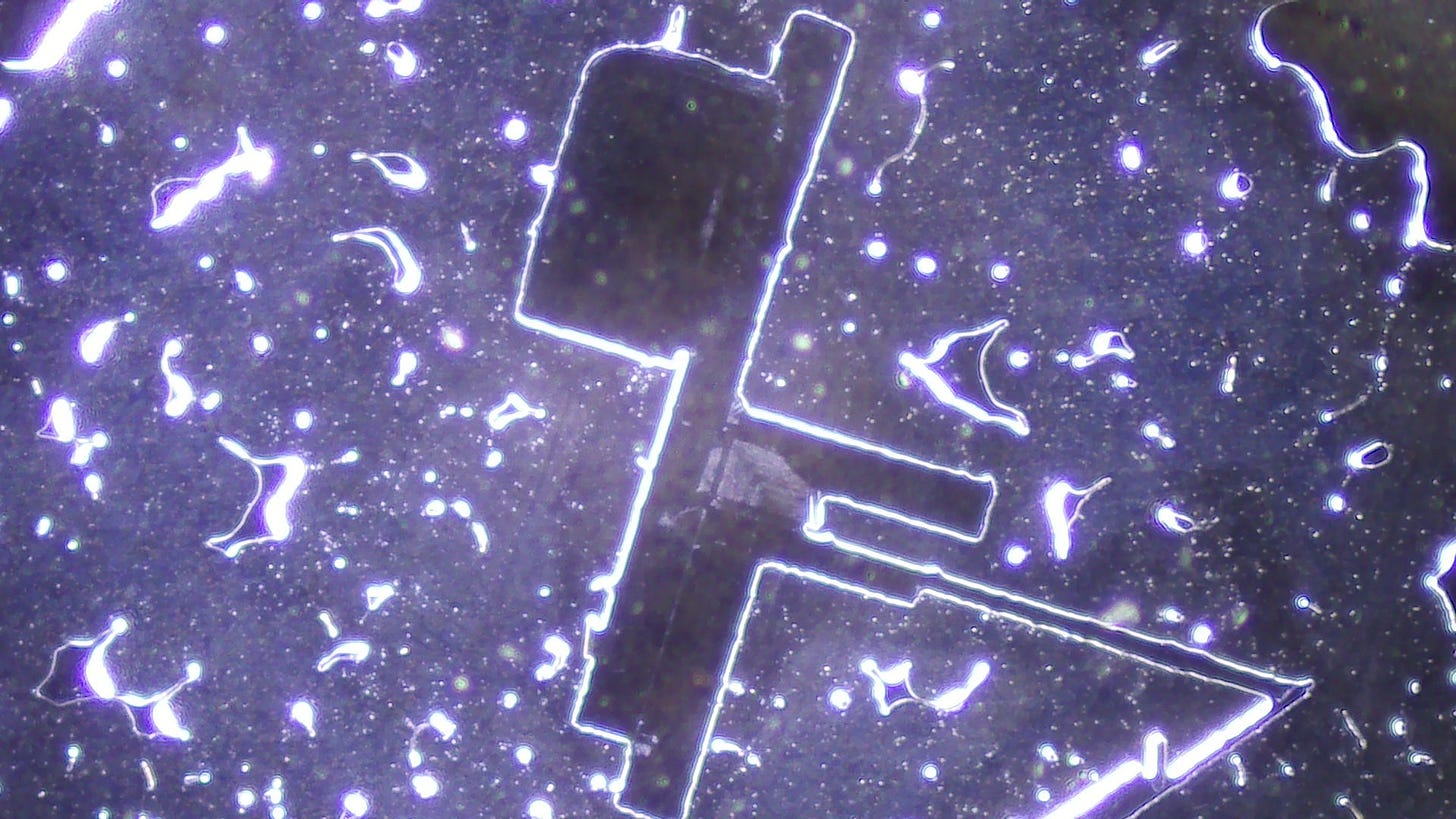
Next, I took a photo through the eyepiece as the screen magnifies everything four times preventing a good overview. We see an assortment of different sizes of crystals which are most likely Zeolite crystals. The manufacturer states that the Zeolite in the product is nanosized. I measured some of the larger and medium size crystals and they are far larger than nano size.
Above at 40x magnification
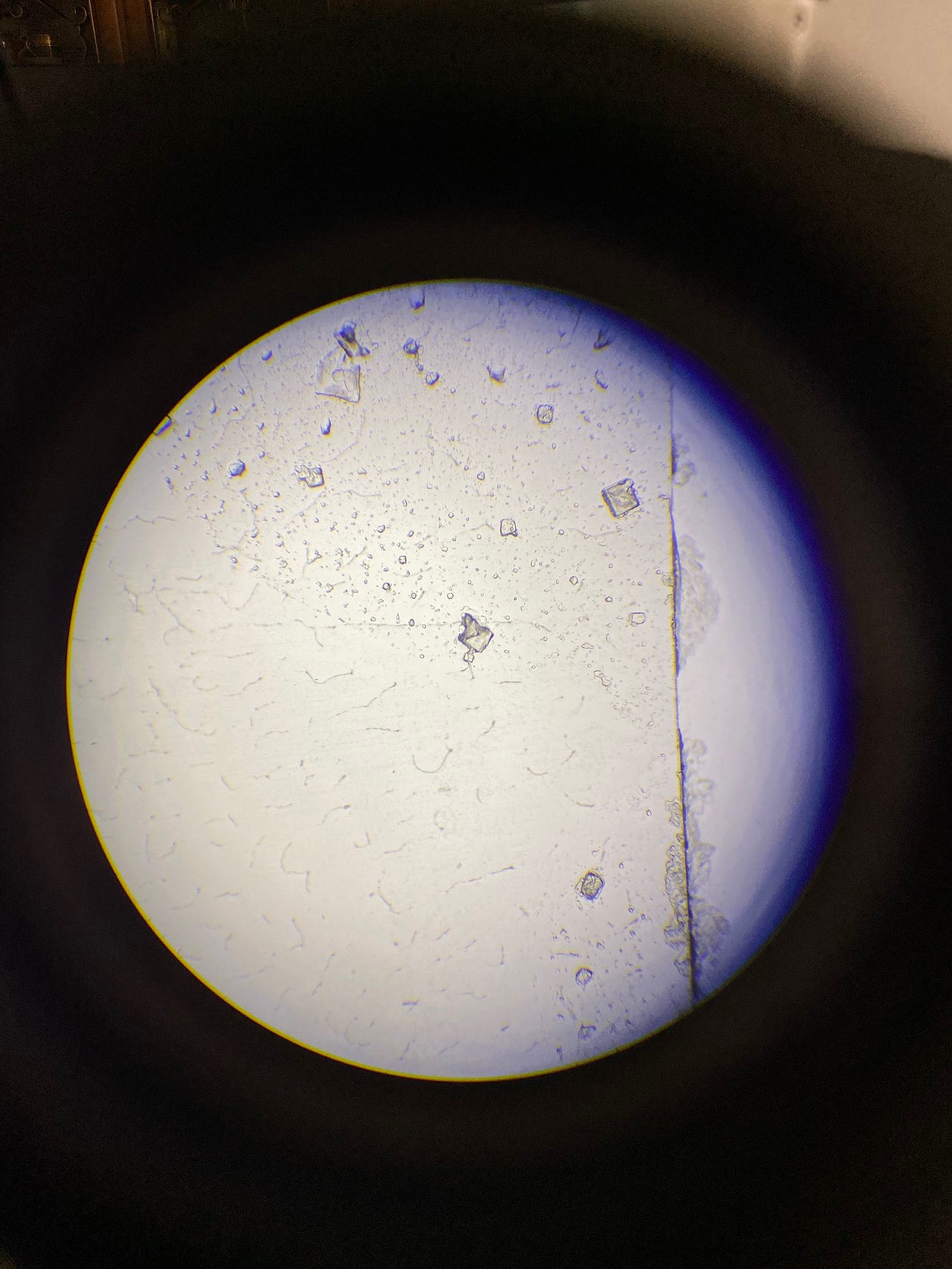
Above at 100 x magnification
Above are the crystals seen through the eye pieces at 40x and 100 x magnification.
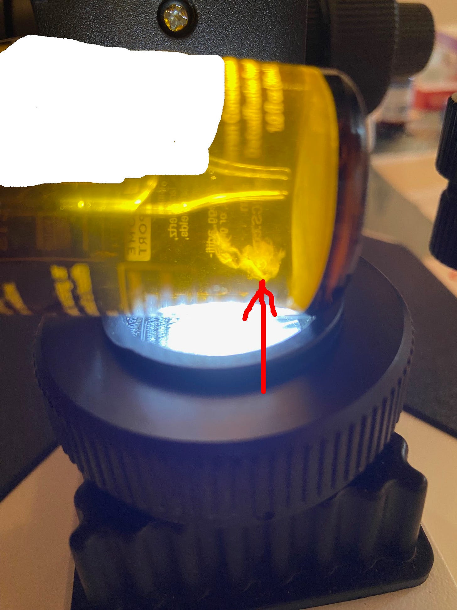
I also took a photo of the bottle which shows things floating around:
I had ordered two bottles. The bottle above is the first bottle I had ordered.
The one below is the second bottle:
I have covered the name of the product as I don’t want to reveal it just yet to prevent a gag order before I publish parts 2 and 3.
Nano sized???
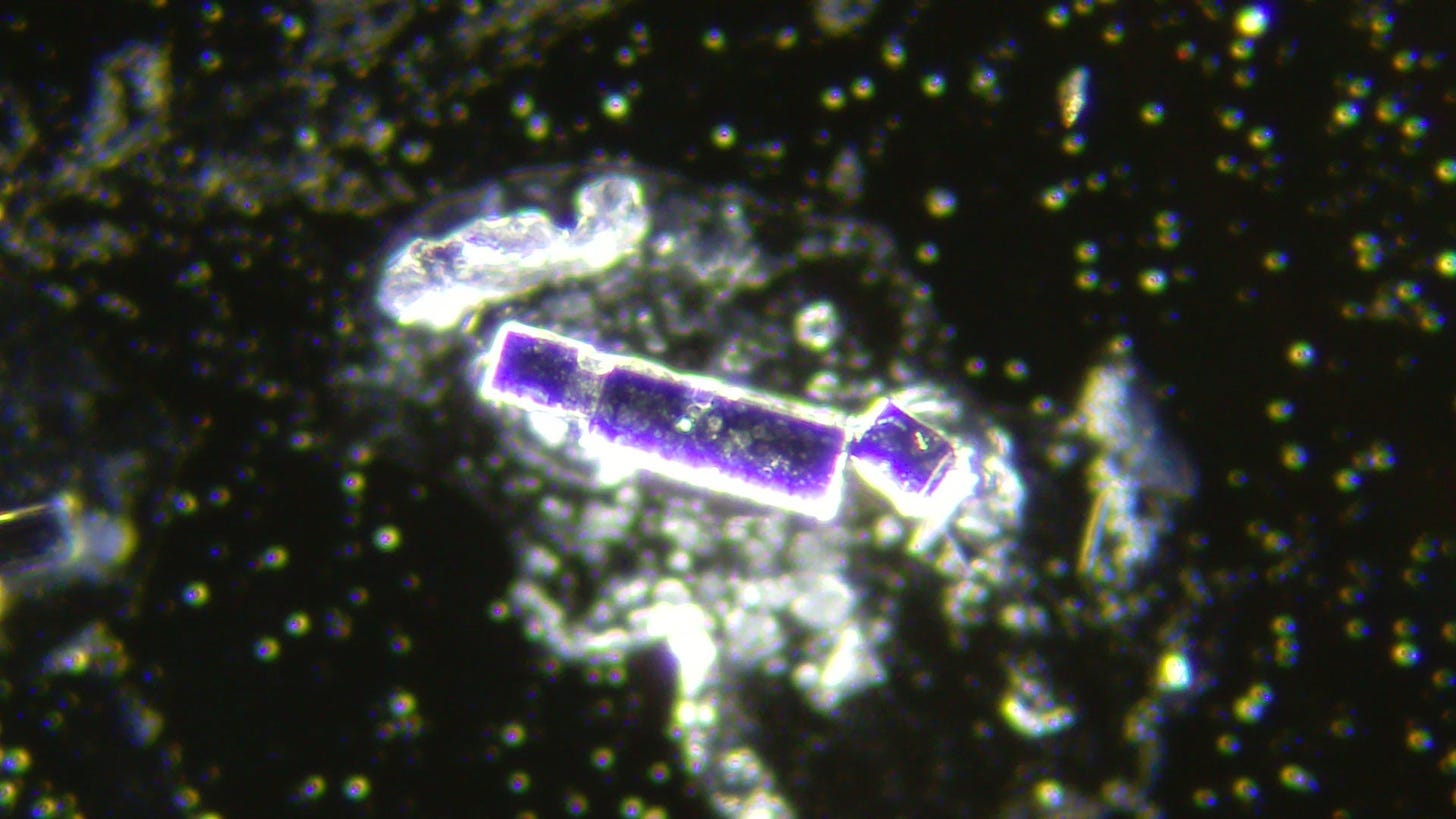
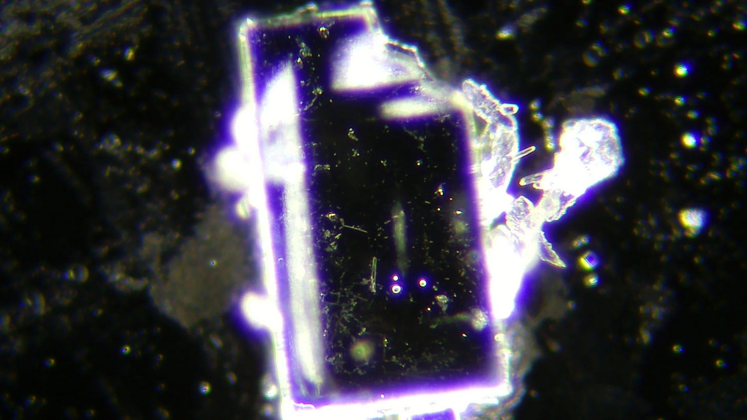
I measured some of the larger crystals. This larger crystal below has a length of 25.4 mm on screen divided by 800 (the magnification) is 0.031 mm which is 31000 nanometers! That isn’t exactly nanosized as claimed by the manufacturer.
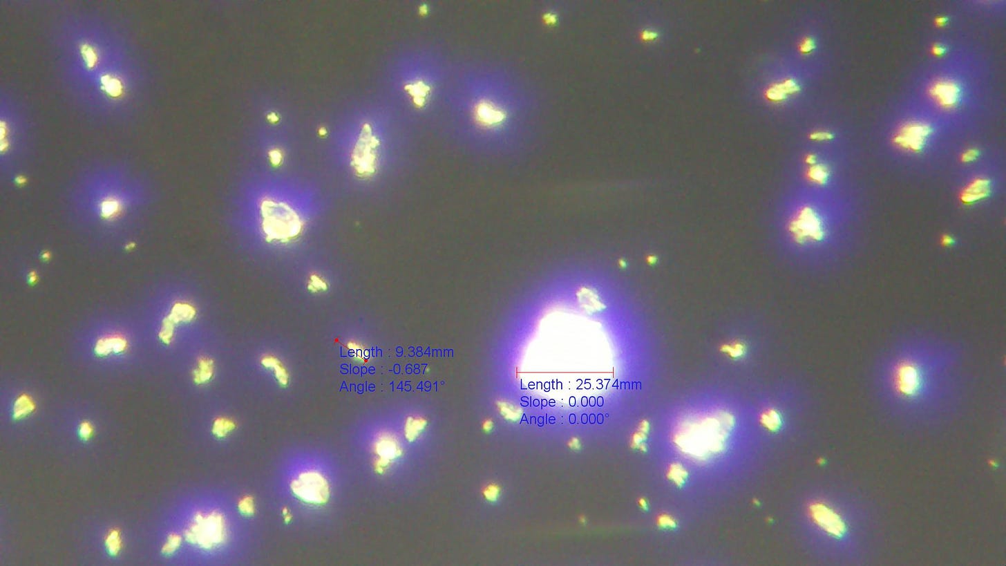
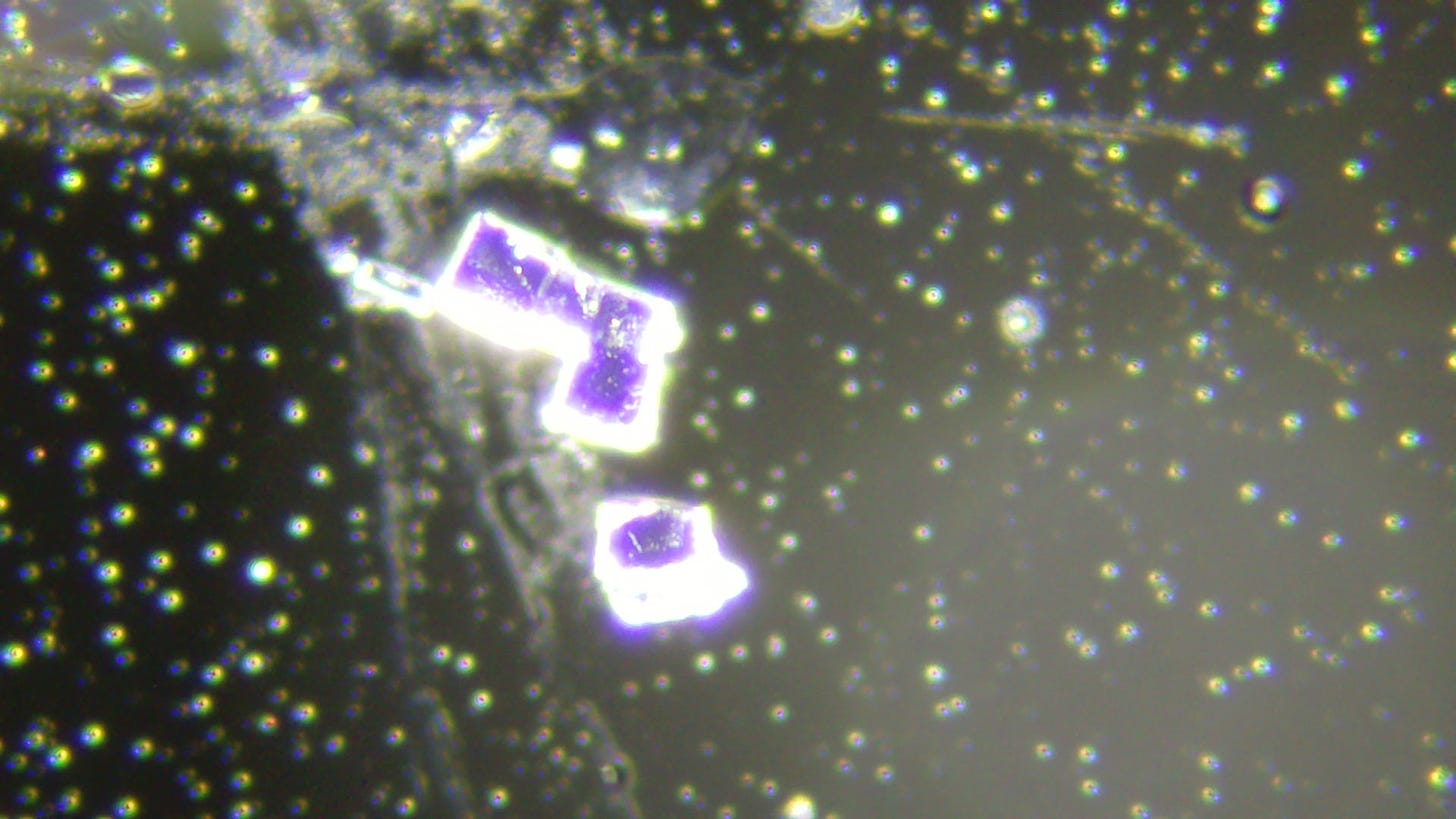
I measured two other crystals at 1600 x magnification. The larger crystal measured 25.37 mm on screen divided by 1600 x magnification is 0.0158 mm in actuality or
15 800 nanometers. The smaller crystal measured 9.384 mm on screen, divided by 1600 comes to 0.00586 mm or 5860 nm.
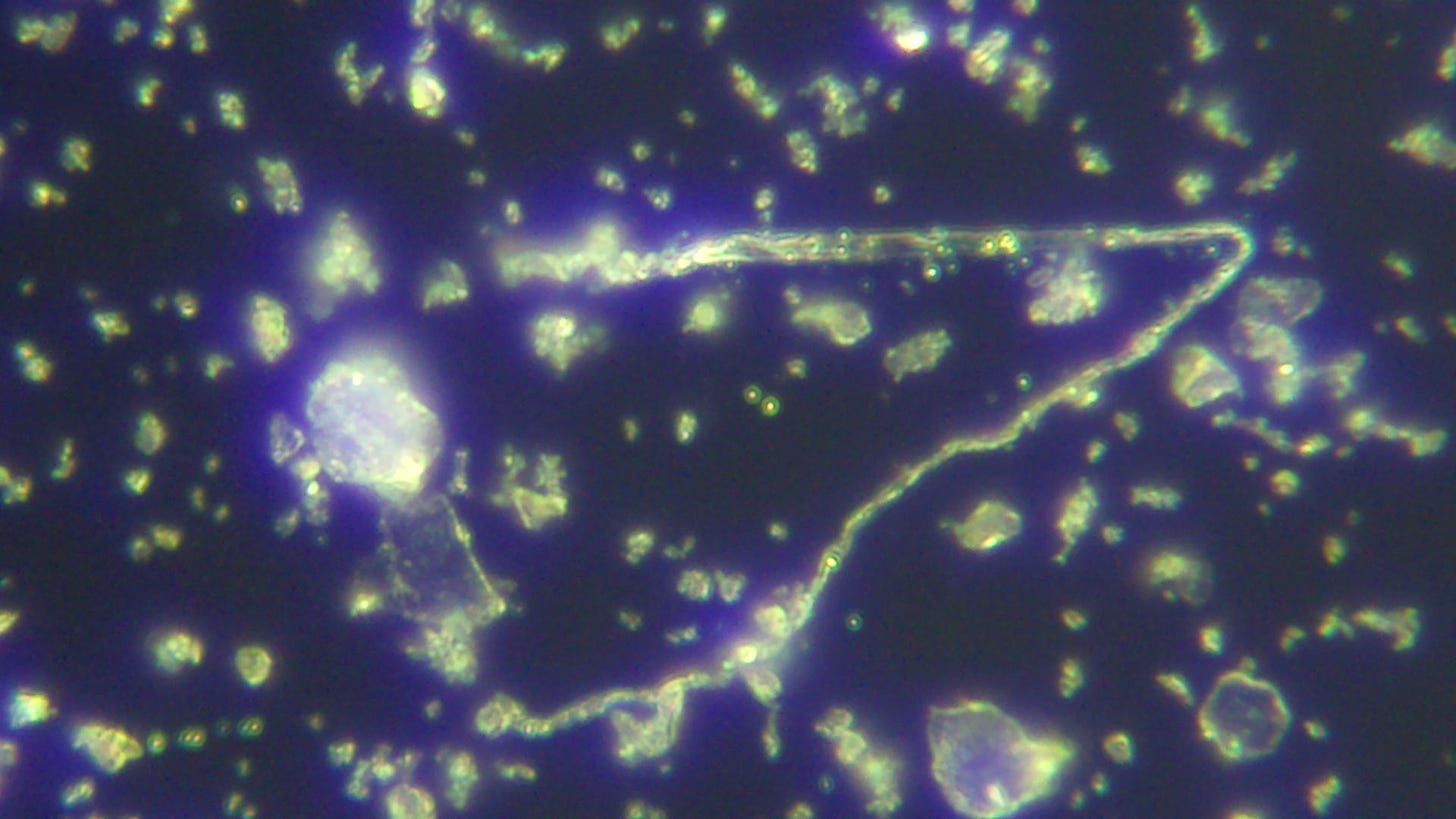
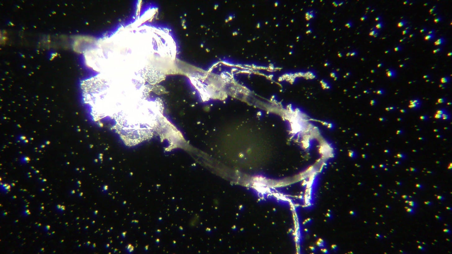
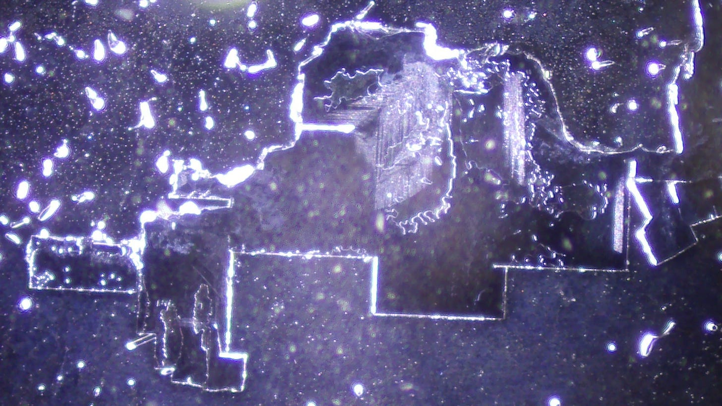
I found some structures that didn’t look like Zeolite crystals:
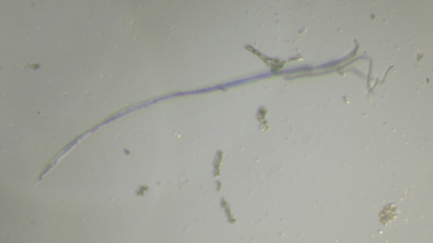
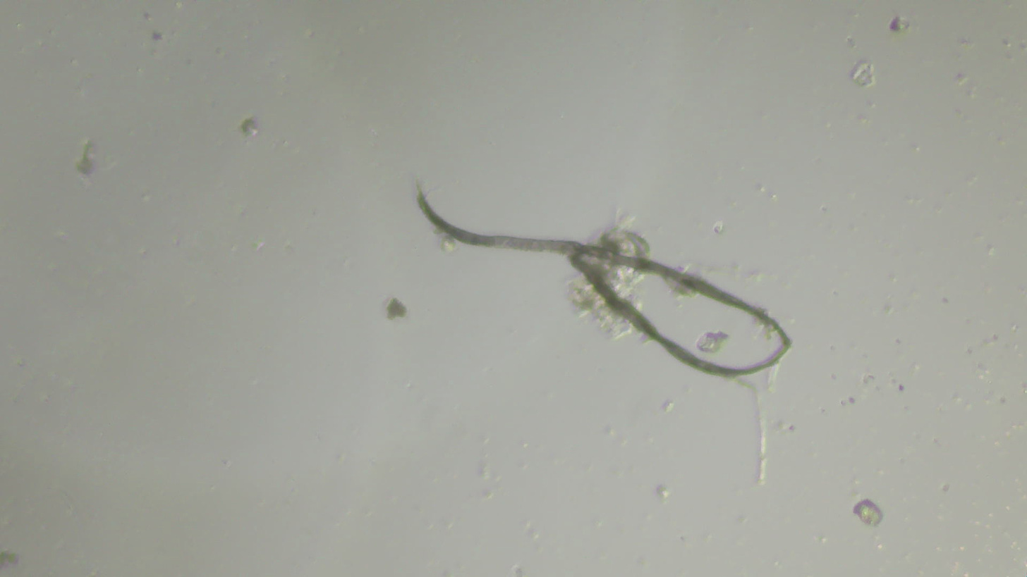
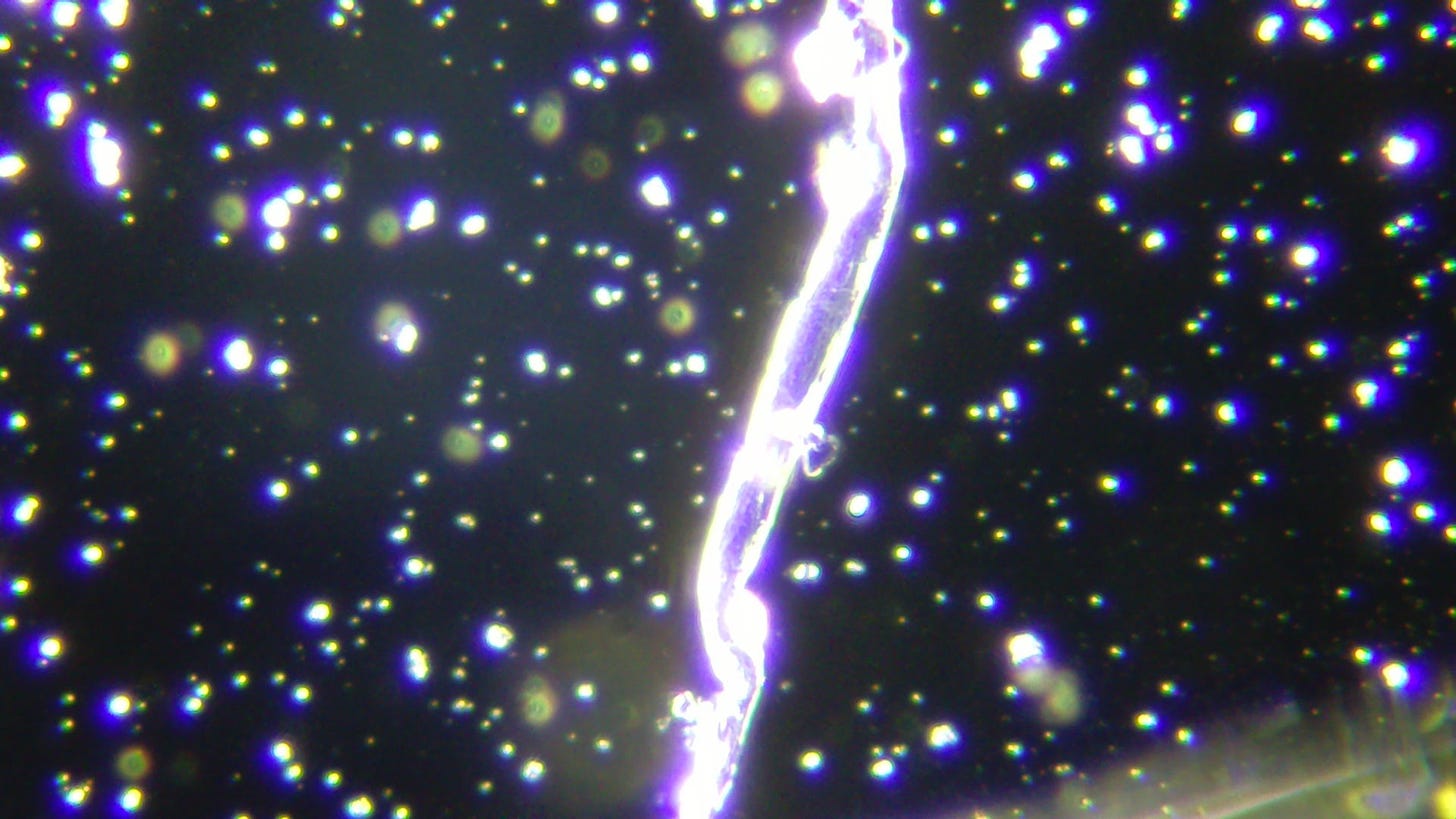
The first image shows a fiber that’s not supposed to be there, and it isn’t an artifact.
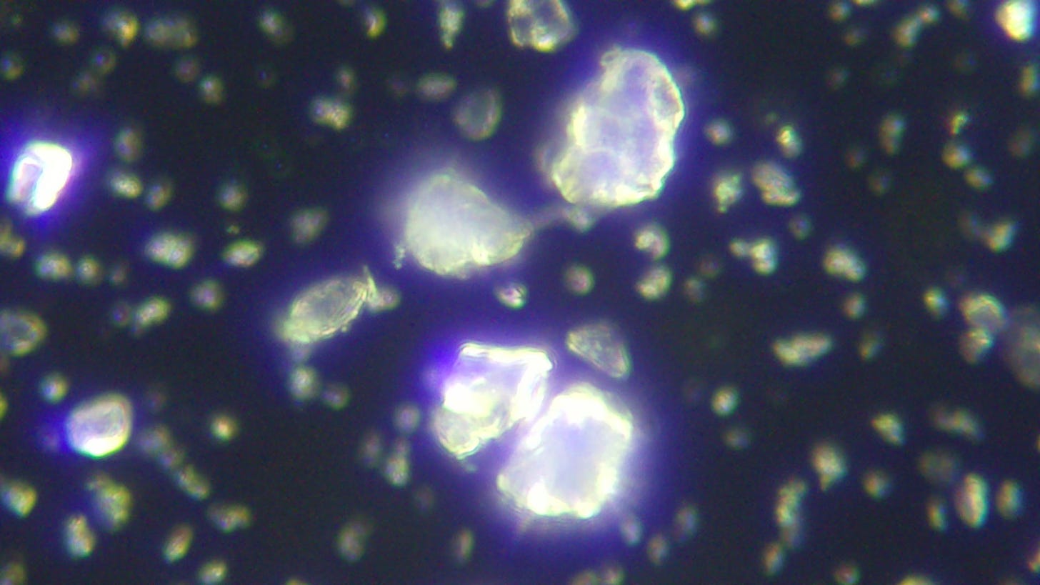
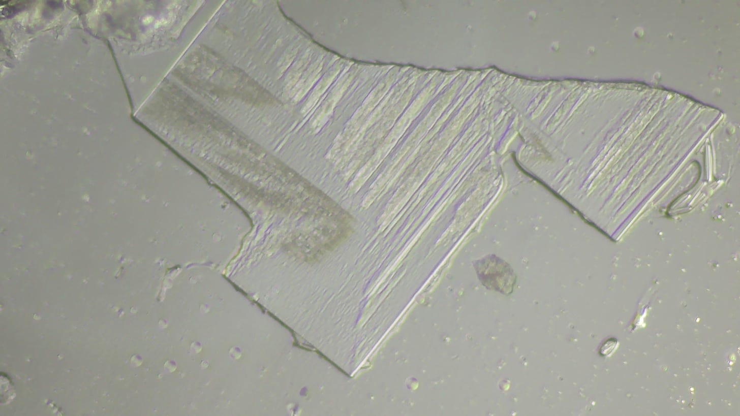
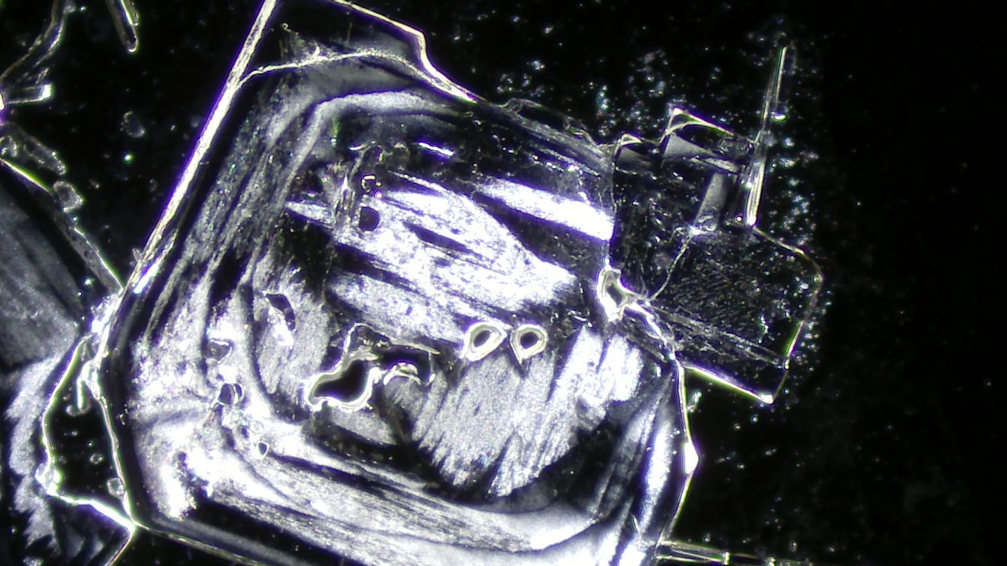
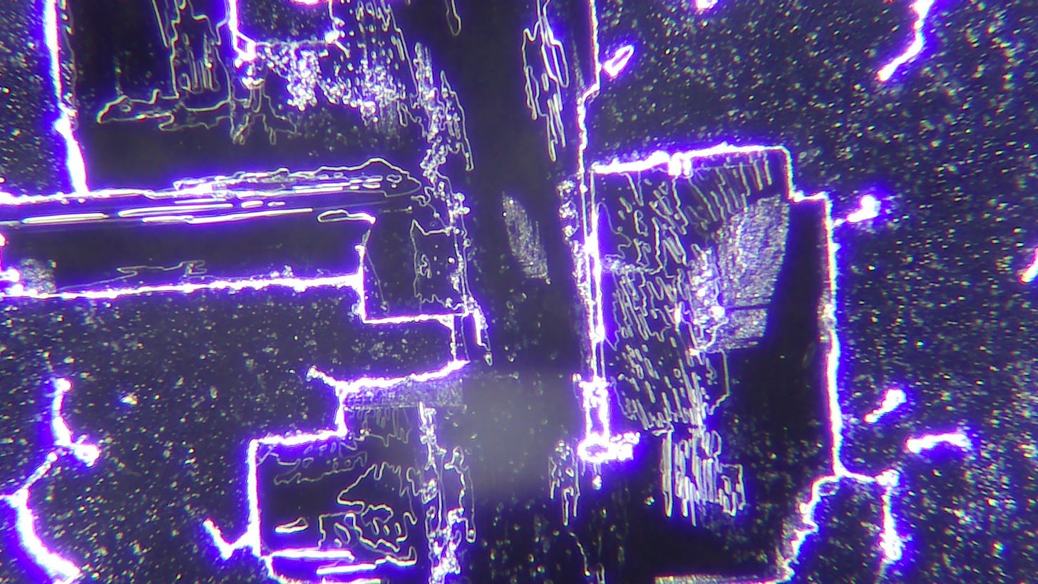
The first photo below shows Zeolite crystals of various sizes but the photo below that at 4000 magnification shows a structure that looks different from the regular Zeolite crystals.
This last structure looks similar to hydrogels or possibly even Graphene Oxide as I have been taught by COV-ID’s Substack who is part of La Quinta Columna. Ariyana Love agrees with me that it looks like a hydrogel, see her comment:
“Dr. Ariyana Love (ND) commented on your post Masterpiece of Deception- A Trilogy.
It's obvious what supplement you're testing here. I'd like to see your urine sample without any added substance, as a control. Why didn't you show this? How do we know the strange structures were not already there in your urine, since they're showing up in almost everybody these days? The image of what you said looks like "graphene", looks more like a polymer hydrogel fiber….”
She thought I did microscopy on a mix of urine and the product which I did not. The only thing under the microscope was the product.
I wanted to see what happens if the slide is exposed to EMF. I put a slide with a drop of the product on a wireless phone charger for 70 hours.
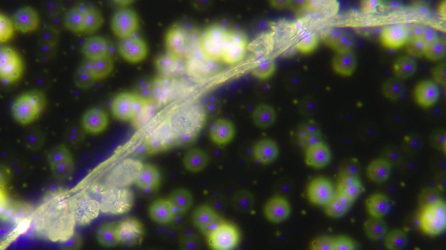
In bright field I found more fibers and shapes with sharp edges and corners.
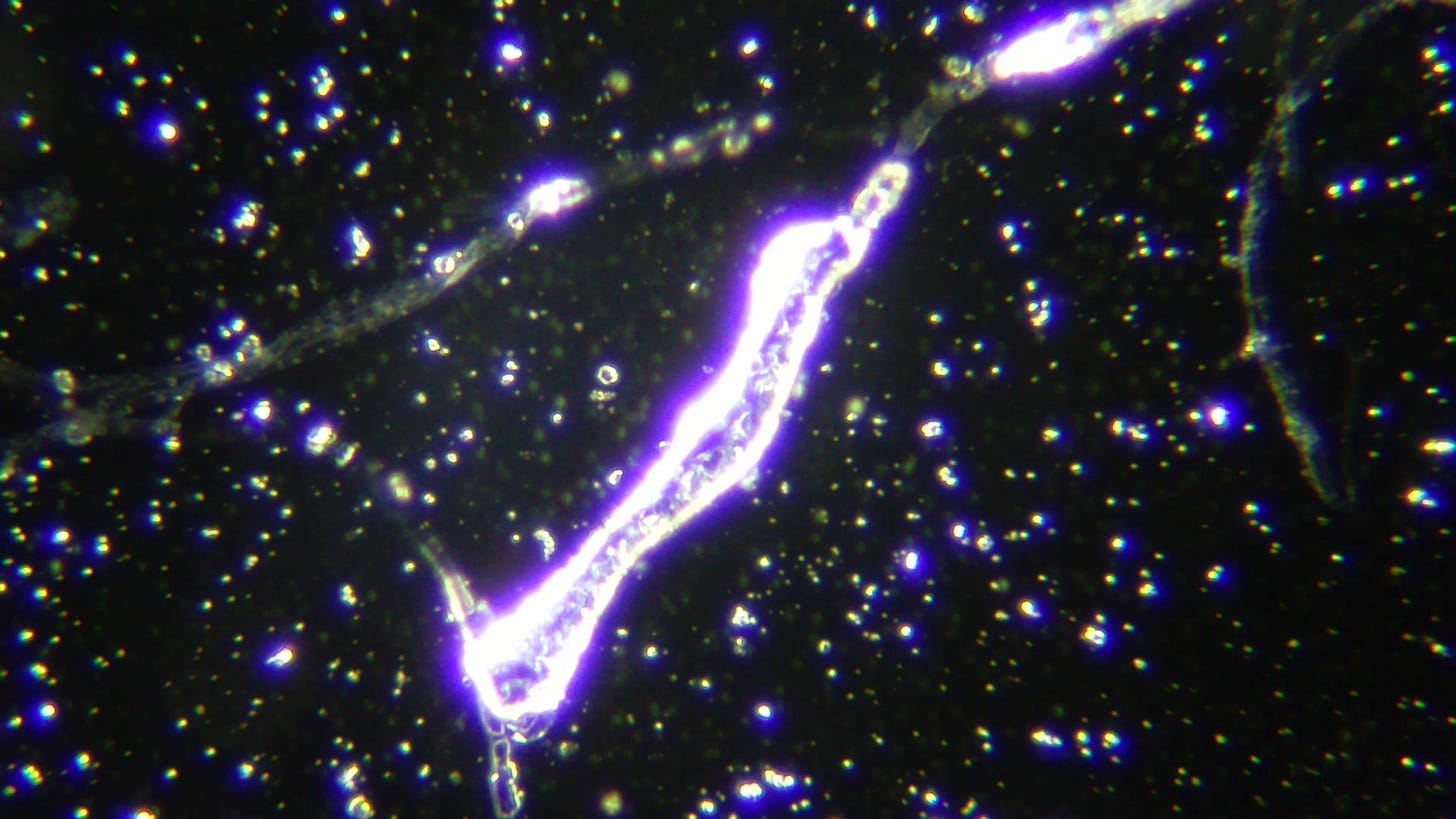
In dark field many more fibers showed up and undefined structures that didn’t look like Zeolite crystals.
Twelve Days on EMF during the Tornado/Hurricane Intermezzo
I placed the slide back on the wireless charger and then forgot about it as I was busy with a tornado in a hurricane whirling by my house. After I restored my house from the storm, I discovered that I still had the slide on the charger pad. Twelve days had gone by. I was curious to see if some other stuff grew and wasn’t disappointed.
The following two images were taken through the eye piece at 100x and 40 x magnification.
100 x magnification above and 40 x magnification below
Many huge fibers grew while exposed to EMF.
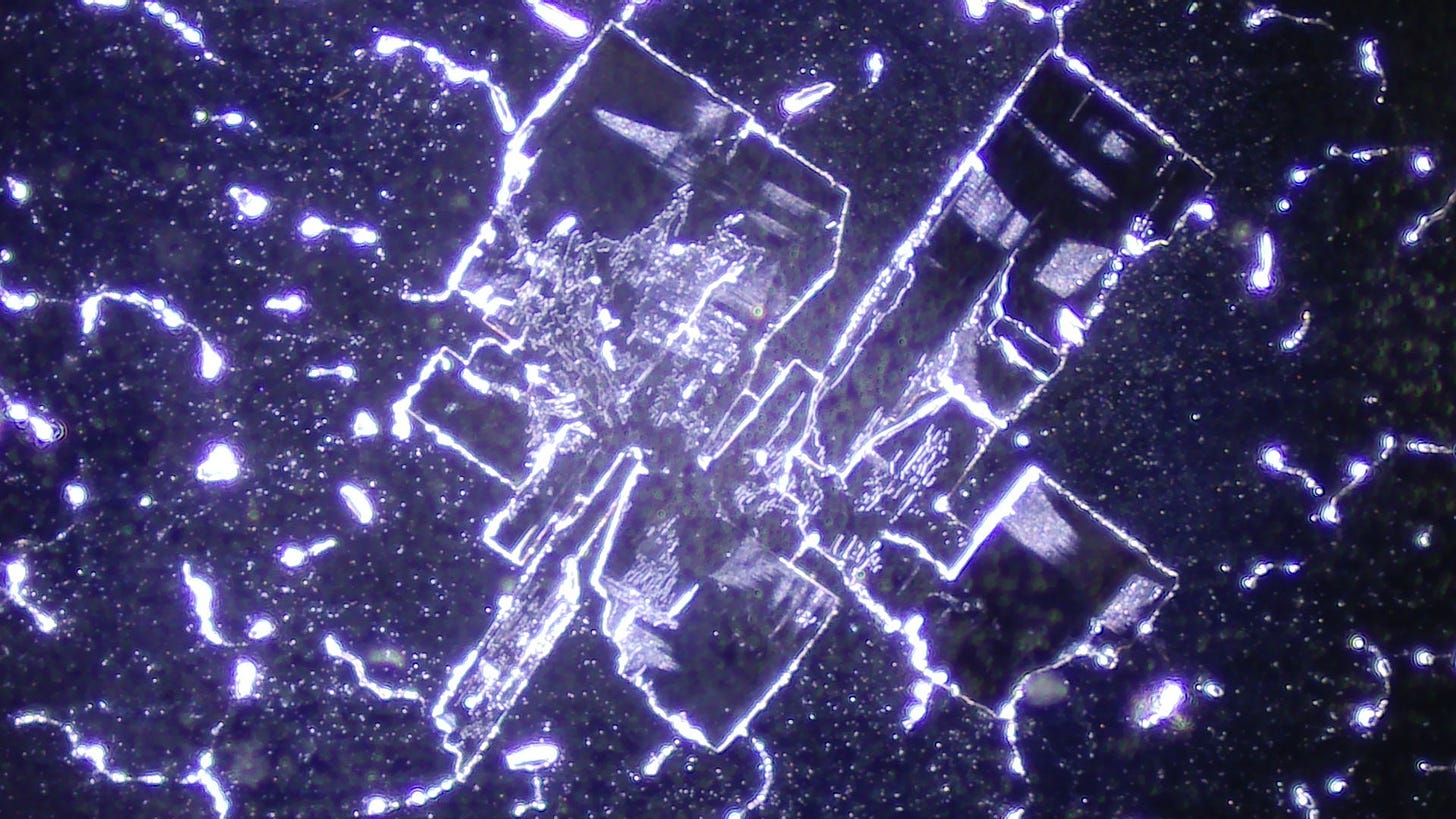
The following images are magnified by 160 to 800 and showed structures that weren’t there before this longer EMF exposure.
160 x magnification
160 x magnification
400 x magnification
400 x magnification
400 magnification
800 x magnification
These are new structures that certainly don’t look like zeolite crystals, but more like computer chips and they are fairly large as the magnification was only 160 -400 except for the last picture. They could be salt crystals as the product contains Sodium and other minerals. To see if they appear consistently around the edges of the coverslip where it would begin the drying process first, I put a drop of this product on a slide on the phone charger pad for 4 days and looked at it in bright field. It showed square structures around some of the edges of the cover slip. When the sample begins to dry it begins at the outer edges of the cover slip. They should thus consistently appear all around the edges. Strangely, these crystals weren’t distributed all the way around the edges of the cover slip, but only intermittently.
Upper edge of the cover slip with an accumulation of crystals.
Right edge of the cover slip with an accumulation of crystals only in some areas.
The drying process happens all around the edge, not just in certain places. So that puts a question mark behind the crystal theory as they only appear in certain areas around the edges.
I checked a slide with blood I had put on the phone charger for the same amount of time and the blood dried quite evenly all around the edges of the cover slip as the following video shows.
The black ropey structures are Fibrin in dried blood.
See similar structures found by Dr. David Nixon here:
I checked it in dark field as well, and as Dr. David Nixon wrote in his post above, it doesn’t disappoint:
I have been looking through microscopes since 1986 and I’m a certified by The Complementary Medical Association as a Live Blood Analyst.
In the next part of this trilogy, I will focus on studies showing the use of zeolite to enhance nanotechnology. And we’ll see if the addition of this product to jabbed blood makes any difference.
Note about your comments: I read them all and I appreciate your questions and suggestions. Unfortunately, the comments have been taking up more and more of my limited time which is why I decided not to respond to all of them any more as I’m doing this research besides my regular work (the one that pays the bills), advising the detox clinic (for free) that will open in about a week and taking care of my little sister who has Down Syndrome as well as my household, vegetable garden and planting season just began.
Book Store: www.anitabaxasmd.com


